


The upper limb is a highly mobile structure designed for precision, grasping, and fine motor manipulation. To support these complex movements, the limb requires a robust vascular network to deliver oxygenated blood and a sophisticated drainage system to remove metabolic waste and manage interstitial fluid. Understanding the transition of vessels through key anatomical landmarks—such as the axilla, the cubital fossa, and the carpal tunnel—is essential for clinical practice and surgical anatomy.
Arterial Supply: From Trunk to Fingertips
The arterial supply of the upper limb follows a continuous pathway, with the name of the vessel changing as it crosses specific anatomical boundaries.
The Proximal Segment: Subclavian and Axillary Arteries
The journey begins with the subclavian artery. As this vessel crosses the lateral border of the first rib, it changes its name to the axillary artery. The axillary artery is responsible for supplying the shoulder and pectoral regions.
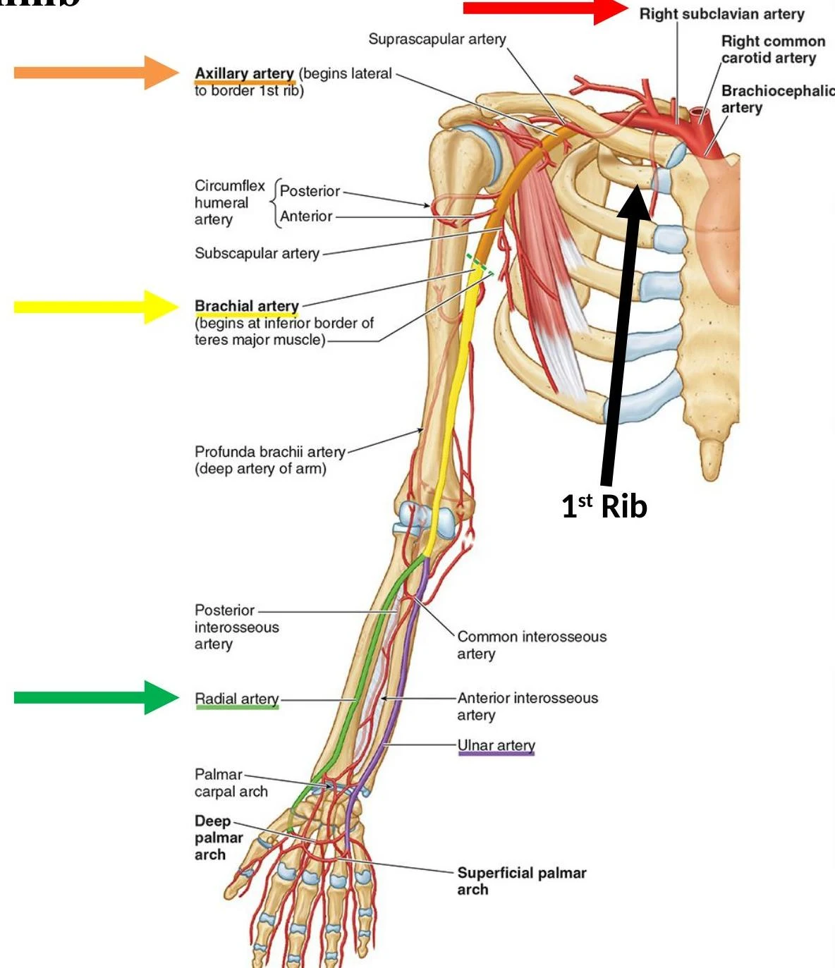
The Arm and Forearm: Brachial, Radial, and Ulnar Arteries
Once the axillary artery crosses the inferior border of the teres major muscle, it becomes the brachial artery. The brachial artery travels down the arm and enters the cubital fossa (the triangular depression anterior to the elbow), where it eventually terminates by dividing into two main branches:
- Radial Artery: Located on the lateral aspect of the forearm.
- Ulnar Artery: Located on the medial aspect of the forearm.
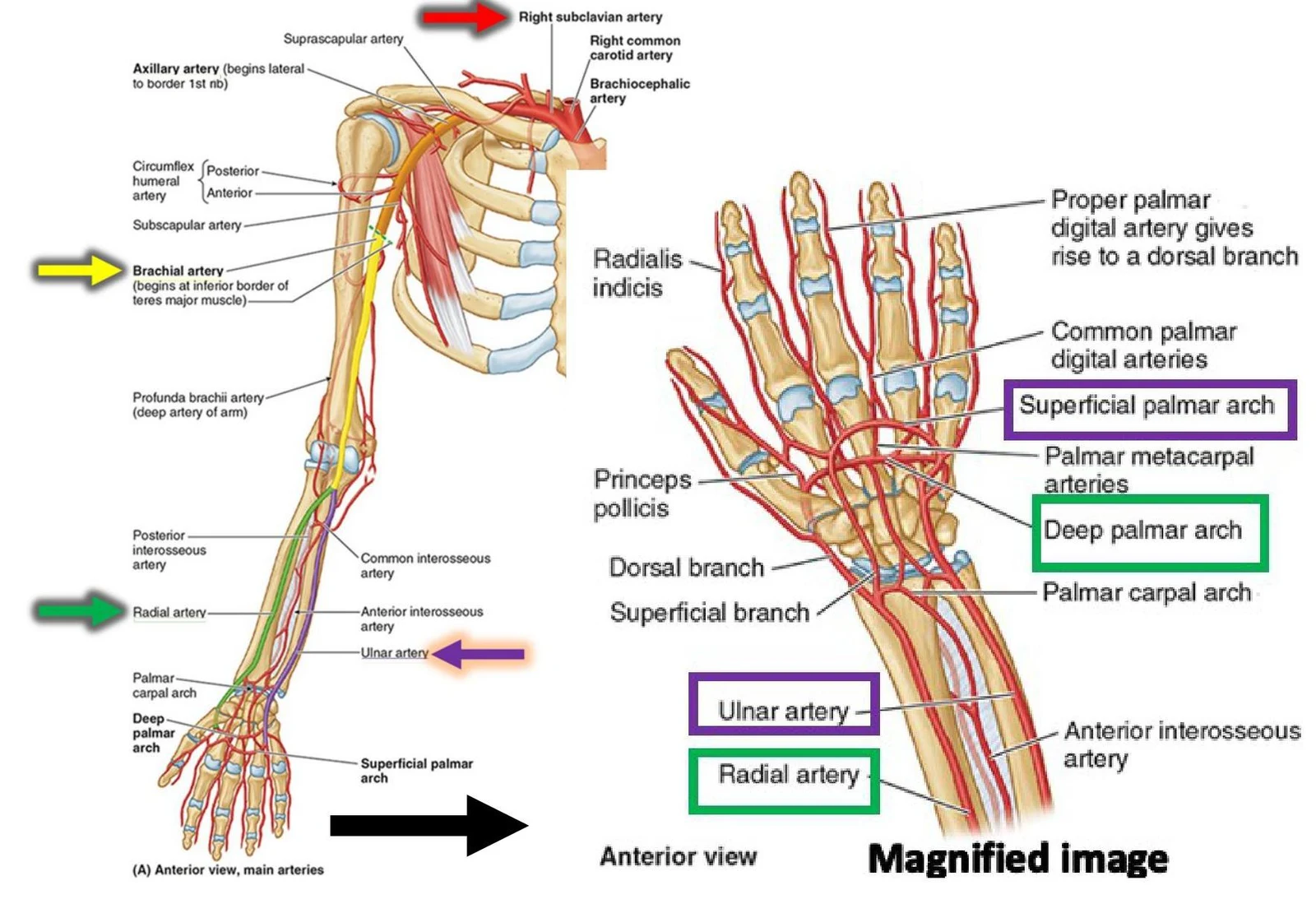
The Hand: Palmar Arches
As the radial and ulnar arteries reach the hand, they do not simply end; they communicate through a network of anastomoses known as the palmar arches. Specifically, the ulnar and radial arteries typically connect to form the superficial palmar arch and the deep palmar arch.
Keep reading for free.
Create an account to finish this reading and make it part of your study routine.
Continue reading freeAlready have an account? Log in
Updated