An anatomical overview of the scalp layers, neurovascular supply, and the protective meningeal membranes of the brain and spinal cord. Includes clinical correlations related to pain and cerebrospinal fluid dynamics.
The human central nervous system is protected by a sophisticated multi-layered defense system. This protection begins externally with the scalp, a highly vascularized and innervated structure, and continues internally with the meninges—three specialized membranes that cushion the brain and spinal cord. Understanding these layers is essential for clinical practice, particularly when managing head trauma, infections, or intracranial pressure.
The Scalp: Layers and Structure
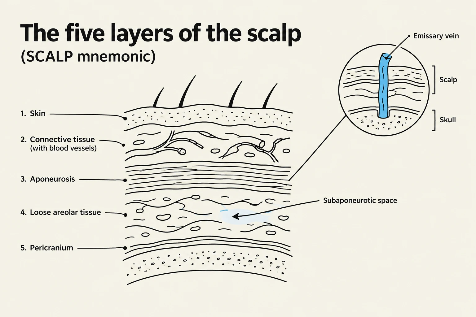
Diagram illustrating the five layers of the scalp: skin, connective tissue, aponeurosis, loose areolar tissue, and pericranium, with a magnified view of an emissary vein.
The scalp is not a single unit but a complex arrangement of five distinct layers. A helpful mnemonic to remember these layers is the acronym SCALP:
S - Skin: Thick, hair-bearing, and rich in sebaceous glands.
C - Connective Tissue: A dense, fibrofatty layer containing numerous blood vessels.
A - Aponeurosis (Epicranial): A thin, tendinous sheet (the epicranial aponeurosis) that connects the frontal and occipital muscles.
L - Loose Areolar Tissue: This layer occupies the subaponeurotic space, a potential space located between the epicranial aponeurosis and the underlying pericranium (the periosteum of the skull).
P - Pericranium: The periosteum covering the outer surface of the skull bones.
Clinical Pearl: The Subaponeurotic Space
The loose areolar tissue (the "L" layer) allows the first three layers of the scalp to slide as a single unit over the skull. However, this space also contains emissary veins. These are valveless veins that connect superficial scalp veins to the intracranial venous sinuses. Because they lack valves, they can provide a direct pathway for infections to travel from the scalp into the cranial cavity.
Neurovascular Supply of the Scalp
The scalp is characterized by its intense vascularity and rich nerve supply, which explains why even minor lacerations can bleed profusely.
Arterial and Venous Supply
The arteries typically run through the dense connective tissue layer, following the paths of the nerves. Key arteries include:
Supratrochlear and Supraorbital arteries: Branches of the ophthalmic artery (internal carotid).
Superficial Temporal artery: A terminal branch of the external carotid.
Occipital artery: A branch of the external carotid supplying the back of the scalp.
Venous drainage involves the facial vein, the retromandibular vein, and the external jugular vein. As mentioned, the emissary veins link these superficial vessels to the internal dural venous sinuses.
Sensory Innervation
The sensory supply is divided into two main groups based on their location relative to the ear:
Anterior to the ear (Trigeminal Nerve): Includes the supratrochlear, supraorbital, zygomaticotemporal, and auriculotemporal nerves.
Posterior to the ear (Cervical Spinal Nerves): Includes the lesser occipital (C2) and the greater occipital nerves.
The Meninges: Protective Membranes
Diagram illustrating the layers of the meninges and the subarachnoid space surrounding the brain.
The meninges are three protective membranes derived from the neural crest and mesenchyme that surround the brain and spinal cord.
1. Dura Mater (Pachymeninx)
The dura mater is the outermost, thickest, and toughest layer. In the cranium, it consists of two distinct layers:
Endosteal layer: The periosteum covering the inner surface of the skull bones.
Meningeal layer: The "dura mater proper," a dense fibrous membrane.
The two layers are closely united except where they separate to form the dural venous sinuses. Unlike the cranial dura, the spinal dura mater lacks the periosteal layer.
Dural Foldings
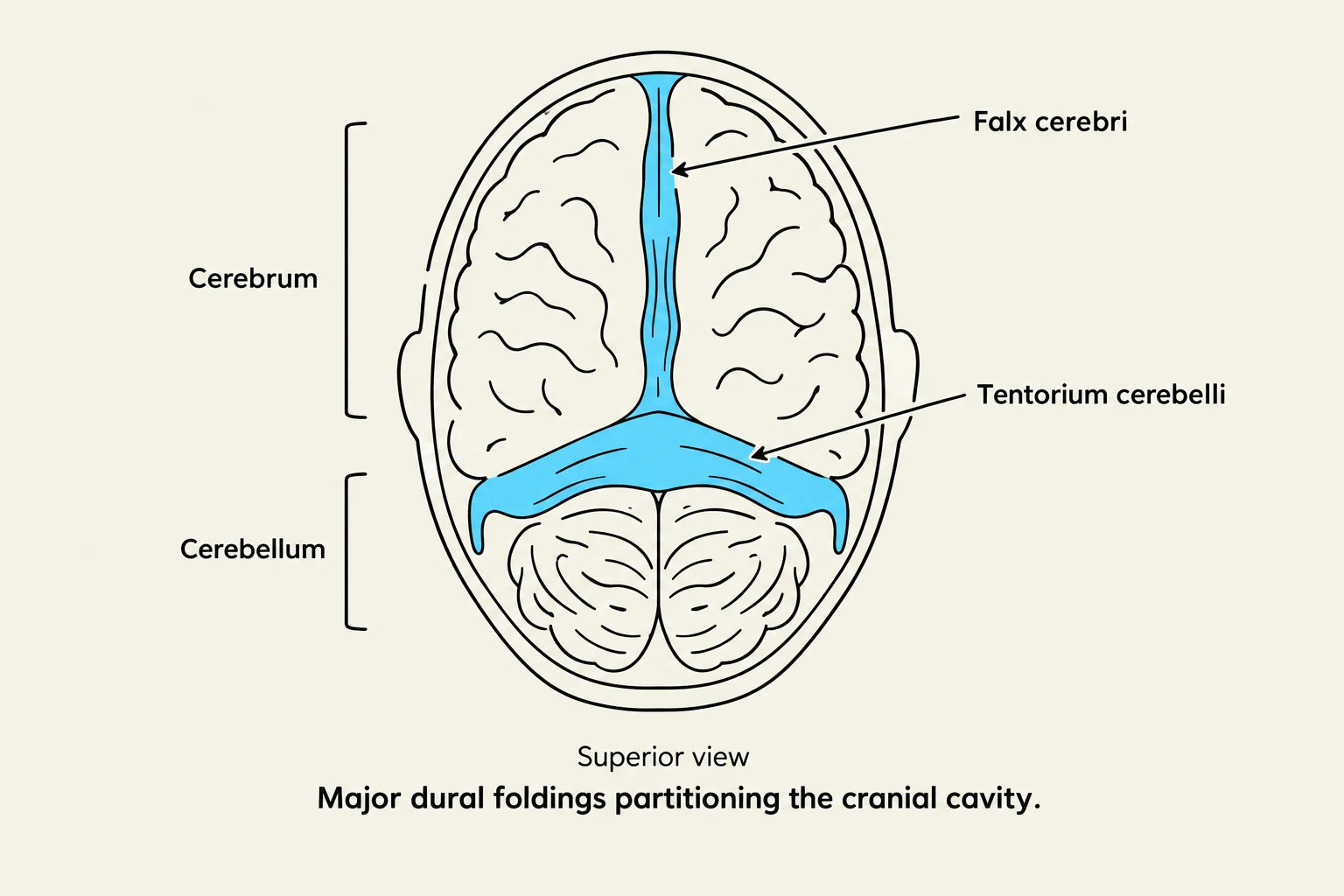
The dura mater forms several significant inward reflections or foldings that partition the cranial cavity:
Falx cerebri: The largest folding; it sits in the midline and separates the two cerebral hemispheres.
Tentorium cerebelli: Separates the supratentorial compartment (cerebrum) from the infratentorial compartment (cerebellum and midbrain).
Falx cerebelli: A smaller folding that separates the two cerebellar hemispheres.
Diaphragma sella: The smallest folding; it forms the roof of the hypophyseal fossa and encircles the pituitary stalk.
2. Arachnoid Mater
The middle layer is a delicate, impermeable membrane. It is separated from the dura by the potential subdural space and from the pia mater by the subarachnoid space.
The arachnoid mater projects into the venous sinuses to form arachnoid villi (or arachnoid granulations/Pachon bodies). These structures are vital for the circulation of cerebrospinal fluid (CSF), as they allow CSF to diffuse from the subarachnoid space into the bloodstream.
3. Pia Mater
The innermost layer is the pia mater, a highly vascular membrane that closely invests the brain. Unlike the arachnoid, the pia follows every contour of the brain, descending into the deepest sulci and covering the gyri.
Terminology Note: Leptomeninges
In clinical anatomy, the term leptomeninges refers specifically to the combined arachnoid mater and pia mater.
Superior view of the major dural foldings partitioning the cranial cavity, showing the falx cerebri and tentorium cerebelli.
Clinical Correlations: Pain and Pressure
Dural Nerve Supply and Headache
The dura mater is highly sensitive to mechanical stimuli. It receives sensory innervation from branches of the trigeminal nerve, the vagus nerve, and the first three cervical spinal nerves.
Because the dura is sensitive to stretching, this is the primary mechanism for producing headaches. The location of the pain often follows the nerve supply:
Above the tentorium: Stimulation of trigeminal endings produces referred pain to the same side of the head.
Below the tentorium: Stimulation of dural endings produces pain referred to the back of the neck and the scalp (along the greater occipital nerve distribution).
Cerebrospinal Fluid (CSF) and Hydrocephalus
The subarachnoid space is filled with CSF, which provides mechanical buoyancy, cushions the CNS from trauma, and removes metabolites.
A disturbance in CSF hydrodynamics can lead to hydrocephalus, an abnormal increase in CSF volume within the skull. This can be caused by: