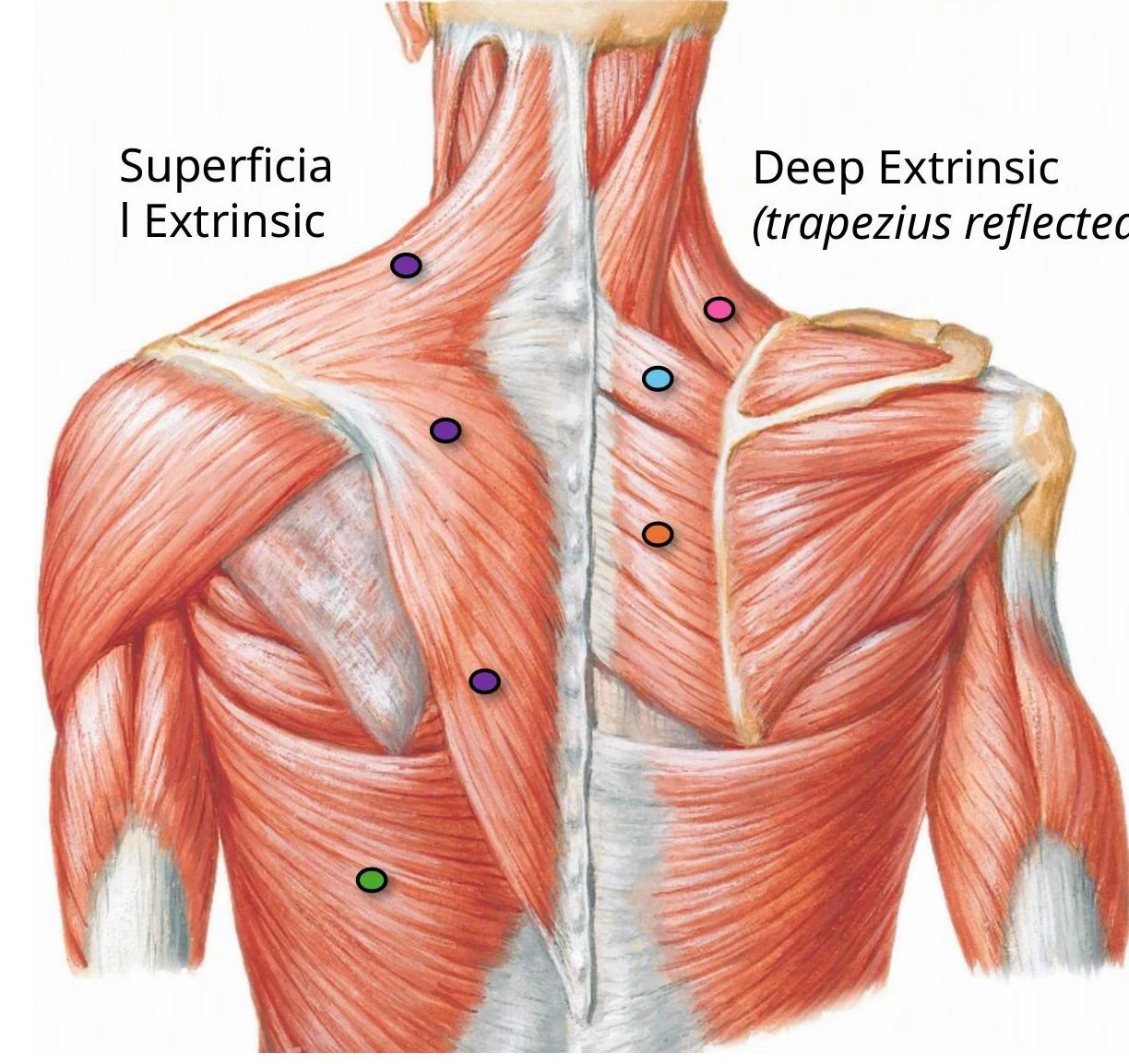
The tendons of these muscles fuse with the fibrous joint capsule to form a musculotendinous cuff that stabilizes the humeral head within the glenoid cavity. Another important muscle in this region is the teres major, which originates from the inferior angle of the scapula and assists in adduction and medial rotation.
Clinical Note: Rotator Cuff Injuries
The supraspinatus is the most commonly ruptured tendon of the rotator cuff. Patients often present with shoulder pain (especially at night), weakness when lifting the arm, and joint crepitus. A "painful arc test" is used to identify supraspinatus tendinitis; a positive test occurs when pain is felt during 60–120° of abduction.
Nerves and Blood Supply
The neurovascular structures of the posterior shoulder must navigate through specific anatomical spaces to reach their targets.
Innervation
- Suprascapular nerve (C5-C6): Courses from the neck through the suprascapular foramen to innervate the supraspinatus and infraspinatus.
- Axillary nerve (C5-C6): Courses from the axilla to the posterior shoulder, passing inferior to the glenohumeral joint to innervate the deltoid and teres minor.
- Subscapular nerves (C5-C6): The upper and lower branches innervate the subscapularis.
Blood Supply
The posterior shoulder benefits from a robust network of anastomoses, known as the scapular arcade, which provides collateral circulation between the subclavian and axillary arteries. Key vessels include:
- Suprascapular artery: Supplies the supraspinous and infraspinous fossae.
- Circumflex scapular artery: Curves around the lateral border of the scapula to enter the infraspinous fossa.
- Posterior circumflex humeral artery: Encircles the surgical neck of the humerus.
- Profunda brachii artery: Passes through the triangular interval.
Gateways to the Posterior Shoulder
The "gateways" are critical anatomical spaces defined by muscle borders. Clinicians must understand these to diagnose nerve impingement or vascular compression.
1. Suprascapular Foramen
This is the route for structures passing from the base of the neck to the posterior shoulder. It is formed by the transverse scapular ligament stretching over the suprascapular notch.
Exam Tip: "Army over Navy"
To remember the contents of the suprascapular notch: the Suprascapular Artery goes over the ligament, while the Suprascapular Nerve goes under the ligament.
2. Quadrangular Space
This space serves as a passage for nerves and vessels between the anterior and posterior shoulder.
- Borders: Superior (teres minor), Inferior (teres major), Medial (long head of triceps), and Lateral (surgical neck of humerus).
- Contents: Axillary nerve and posterior circumflex humeral artery.
Clinical Connection: Quadrangular Space Syndrome (QSS)
Hypertrophy or fibrosis of the muscles surrounding this space can impinge on the axillary nerve or the posterior circumflex humeral artery. This can lead to deltoid weakness, teres minor atrophy, and lateral arm paresthesia.
3. Triangular Space
This area facilitates communication between the axilla and the posterior scapular region.
- Borders: Medial margin of the long head of triceps, inferior margin of teres minor, and superior margin of teres major.
- Contents: Circumflex scapular artery and vein.
4. Triangular Interval
This passage sits between the anterior and posterior compartments of the arm and the axilla.
- Borders: Lateral margin of the long head of triceps, the shaft of the humerus, and the inferior margin of the teres major.
- Contents: Radial nerve, profunda brachii artery, and veins.
Summary Table of Anatomical Gateways
| Space | Key Contents | Clinical Relevance |
|---|
| Suprascapular Foramen | Suprascapular nerve & artery | Nerve impingement at the notch |
| Quadrangular Space | Axillary nerve, Post. circumflex humeral artery | Quadrangular Space Syndrome |
| Triangular Space | Circumflex scapular artery & vein | Scapular collateral circulation |
| Triangular Interval | Radial nerve, Profunda brachii artery | Radial nerve injury |