


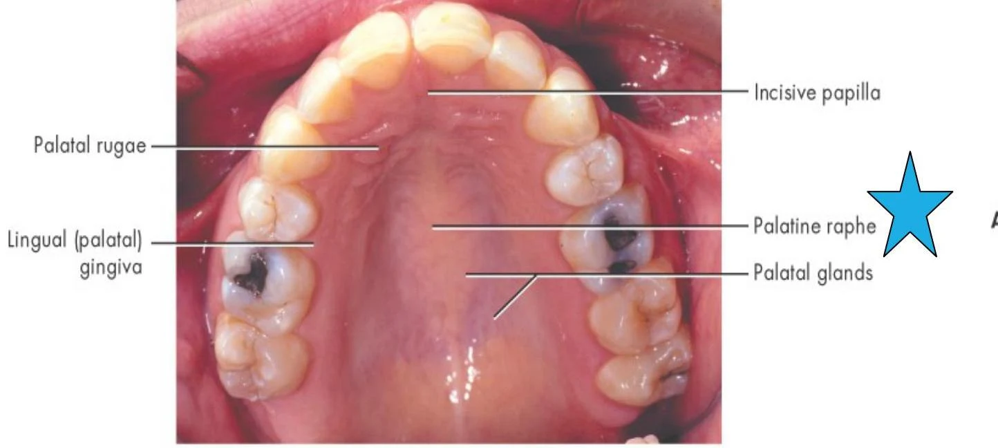
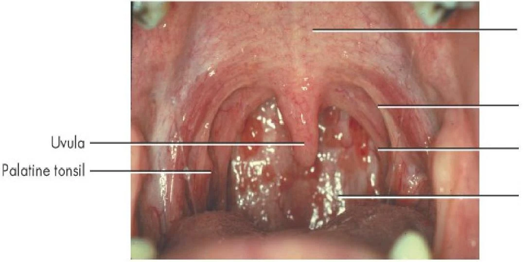
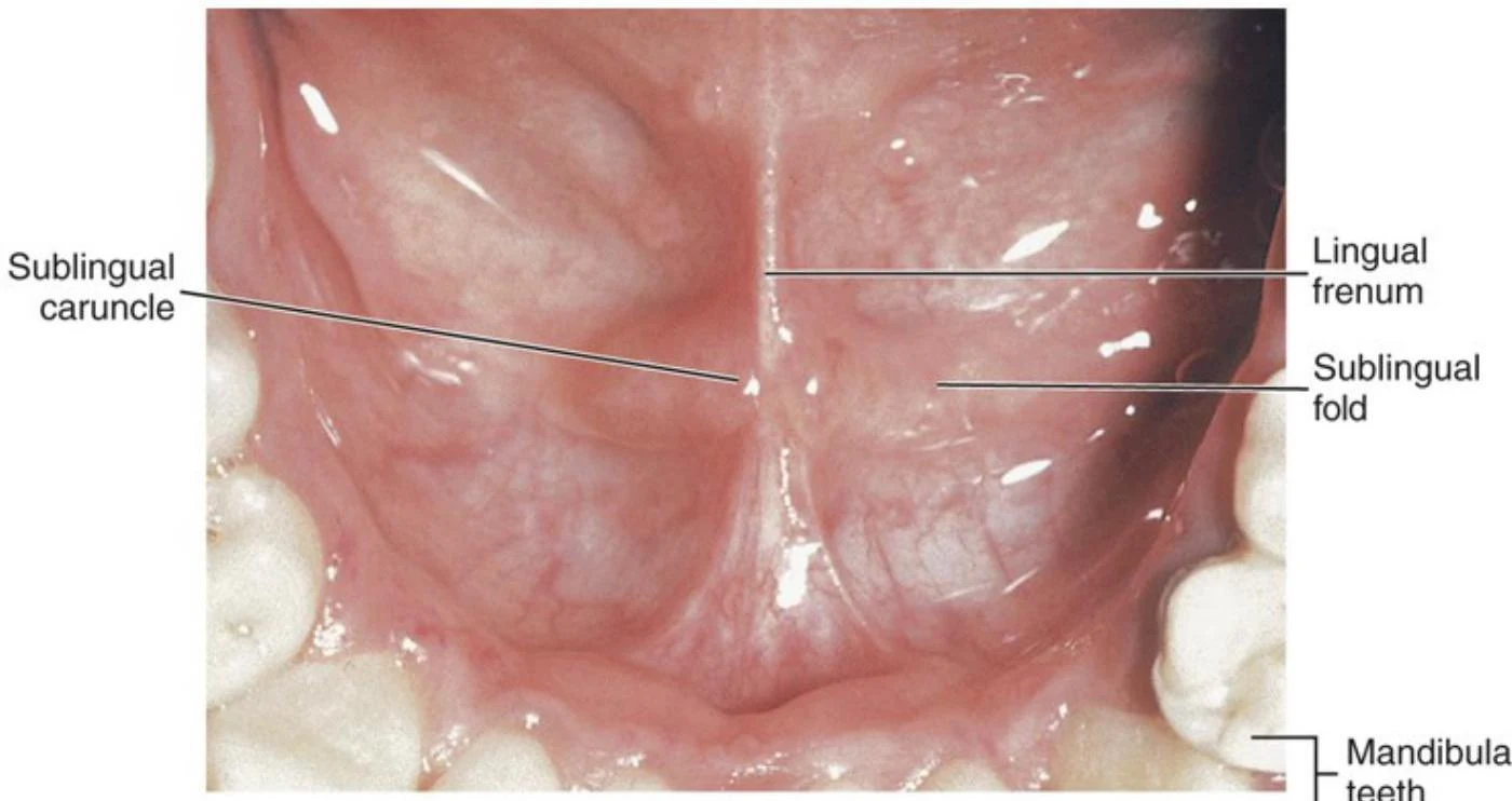
Understanding the intricate anatomy of the oral cavity is fundamental to dental and medical studies. The mouth is not merely a single space but a complex collection of specialized tissues, landmarks, and structures that facilitate functions such as speech, swallowing, and mastication (chewing). This article provides a detailed exploration of the roof of the mouth—the palate—and the various mucosal and anatomical landmarks that define the oral environment.
The Oral Cavity and Mucosa
Before diving into the palate, it is essential to understand the general landscape of the mouth. The oral cavity proper refers to the area located inside the dental arches.

The lining of this cavity is known as the mucosa (mucous membrane). Not all mucosa is created equal; the type of tissue present depends on its location and the amount of physical stress it must endure.
Keratinized Tissue and Masticatory Mucosa
The mouth contains different types of tissue based on their protective needs. Keratinized tissue is tougher and more protective than non-keratinized tissue.
A specific type of tissue called masticatory mucosa is found in areas subject to significant mechanical stress, such as the gingiva (gums) and the hard palate. This tissue is typically keratinized, meaning it has a resilient outer layer to withstand the friction of food during chewing.
In contrast, the alveolar mucosa—which covers the alveolar bone—is a movable, non-keratinized tissue that appears darker pink or red due to its high vascularity. The boundary where the firm, pink attached gingiva meets the looser, redder alveolar mucosa is known as the mucogingival junction.

Keep reading for free.
Create an account to finish this reading and make it part of your study routine.
Continue reading freeAlready have an account? Log in
Updated