


The upper respiratory and digestive tracts begin with a complex arrangement of bony and soft tissue structures designed to facilitate breathing, speech, and swallowing. The nasal cavity serves as the primary gateway for inhaled air, while the palate forms the critical boundary between the respiratory and oral cavities. Understanding these structures is essential for clinical practice, particularly in managing issues such as sinus infections, dental pain, and swallowing difficulties.
The Nasal Cavity: Boundaries and Internal Structure
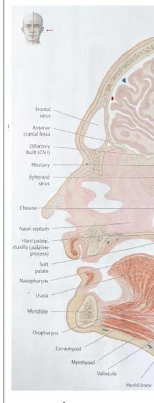
The nasal cavity is a central component of the respiratory tract. It is bounded superiorly by the frontal, ethmoid, and sphenoid bones, which separate it from the cranial cavity. Inferiorly, it is separated from the oral cavity by the hard palate.
The cavity is divided into two symmetrical halves by the nasal septum. This midline partition is composed of three main structures: the perpendicular plate of the ethmoid bone, the vomer, and the septal cartilage.
On the lateral walls of the nasal cavity, three bony projections called conchae (superior, middle, and inferior) extend into the space. These conchae create winding air channels known as meatuses (superior, middle, and inferior meatuses). This complex architecture is not merely for space; it creates air turbulence, which is vital for warming, filtering, and moistening inhaled air before it reaches the lungs.
Clinical Note: Epistaxis Nosebleeds, or epistaxis, often occur due to trauma or dryness at Kiesselbach's plexus, a highly vascularized area located on the anterior part of the nasal septum.
Respiratory Epithelium
The internal surface of the nasal cavity is lined with a specialized tissue known as pseudostratified ciliated columnar epithelium. This lining contains goblet cells, which are responsible for secreting mucus. The mucus serves two primary roles: trapping inhaled dust and pathogens, and moistening the air. The cilia (tiny hair-like projections) then sweep this mucus toward the pharynx to be swallowed, maintaining a clean respiratory pathway.
Keep reading for free.
Create an account to finish this reading and make it part of your study routine.
Continue reading freeAlready have an account? Log in
Updated