Anatomy of the Musculoskeletal System: Bones, Joints, and Muscles
An educational overview of the human musculoskeletal system, covering bone structure, joint mechanics, and muscle function. This guide defines key anatomical terms related to the skeletal and muscular systems, essential for understanding bodily structure and movement.
Understanding the human musculoskeletal system requires a precise vocabulary. Whether you are studying the microscopic layers of compact bone or the complex ways muscles pull on joints to create movement, the terminology provides the blueprint for how the body maintains structure and mobility. This article breaks down these essential terms into three logical categories: bone structure, joint mechanics, and muscle function.
The Architecture of Bone
Bones are not static, dry structures; they are dynamic, living tissues composed of various layers and specialized cells. To understand bone anatomy, we must look at both the macroscopic (visible to the eye) and microscopic (cellular) levels.
At the macroscopic level, long bones consist of an epiphysis (the ends) and a diaphysis (the shaft). The area where these meet is the metaphysis. In growing children, this region contains the epiphyseal plate, a layer of hyaline cartilage that allows for longitudinal growth. Once growth is complete, this cartilage is replaced by bone, leaving behind the epiphyseal line.
Inside the bone, we find different types of marrow. Red bone marrow is responsible for hematopoiesis (blood cell production), while yellow bone marrow serves as a site for fat storage within the medullary cavity. To keep these tissues alive, a nutrient artery foramen allows blood vessels to enter the bone shaft.
Diagram illustrating the structure of a long bone, including the epiphysis, diaphysis, medullary cavity, red marrow, and yellow marrow.
On a microscopic level, compact bone is organized into highly structured units called osteons. These consist of several types of lamellae (layers of bone matrix):
Concentric lamellae: Circular layers surrounding a central canal.
Interstitial lamellae: Remnants of old osteons found between current osteons.
Circumferential lamellae: Layers that wrap around the entire circumference of the bone.
Finally, at the ends of bones where they meet other bones, we find articular cartilage. This smooth, hyaline cartilage reduces friction and absorbs shock during movement.
Exam Tip: When looking at a slide, remember that concentric means "circles within circles," while interstitial refers to the "in-between" spaces.
Joint Anatomy and Movement
Joints (articulations) are the points where two or more bones meet. They are classified by their structure and the amount of movement they allow. While some joints are immovable (fibrous), others are highly mobile (synovial). Cartilaginous joints are a middle ground, where bones are connected by cartilage.
One of the most mobile types of synovial joints is the ball and socket joint, such as the shoulder or hip, which allows for movement in multiple planes.
To describe how we move, anatomists use specific directional terms. These are often categorized by the plane of movement:
Linear and Angular Movements
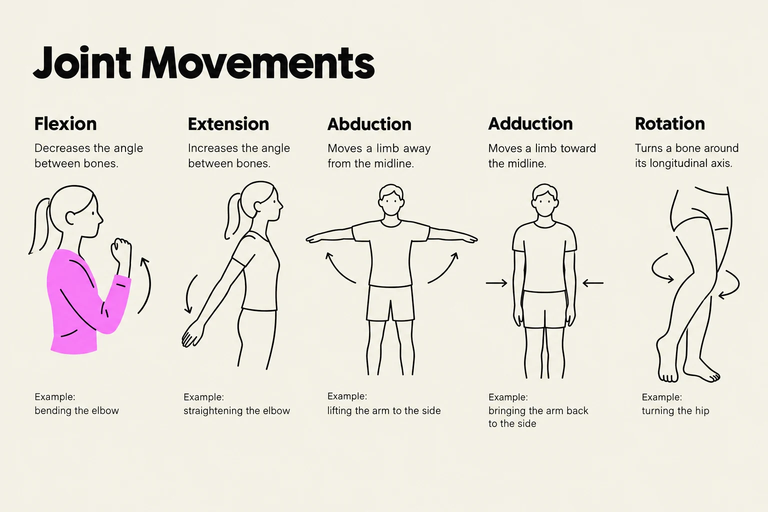
Flexion and Extension: Flexion decreases the angle between bones (e.g., bending the elbow), while extension increases it.
Abduction and Adduction: Abduction moves a limb away from the midline of the body, whereas adduction moves it toward the midline.
Circumduction: A circular movement that combines flexion, extension, abduction, and adduction.
Rotation: Turning a bone around its own longitudinal axis.
Specialized Movements
Elevation and Depression: Moving a body part upward (elevation) or downward (depression), such as shrugging the shoulders.
Protraction and Retraction: Moving a structure forward (protraction) or backward (retraction), commonly used to describe the jaw or scapula.
Supination and Pronation: Specifically used for the forearm; supination turns the palm upward, while pronation turns it downward.
Inversion and Eversion: Used for the foot; inversion turns the sole inward, while eversion turns it outward.
Dorsiflexion and Plantar Flexion: Movements of the ankle; dorsiflexion lifts the toes toward the shin, while plantar flexion points the toes downward.
This illustration demonstrates five types of joint movements: flexion, extension, abduction, adduction, and rotation, with examples for each.
Muscle Anatomy and Action
Muscles are the engines of the skeletal system. A whole muscle is organized into bundles called fascicles, which are further composed of individual muscle fibers (the actual muscle cells). These fibers are wrapped in connective tissue layers: the epimysium (outermost), perimysium (around fascicles), and endomysium (around individual fibers).
Muscles do not work in isolation; they work in functional groups to produce smooth, controlled movements. To describe these roles, we use the following terms:
Agonist (Prime Mover): The muscle that provides the major force for a specific movement.
Antagonist: The muscle that opposes or reverses the movement of the agonist.
Synergist: A muscle that helps the agonist by adding extra force or reducing unnecessary movement.
Fixator: A specialized synergist that stabilizes the origin of the agonist so the movement can occur efficiently.
All these actions occur at specific attachment points, where the muscle's connective tissue is anchored to the bone.
Worked Example: Think of a bicep curl. When you lift a weight toward your shoulder, the biceps brachii is the agonist (it contracts to flex the elbow). The triceps brachii on the back of your arm is the antagonist (it must relax/lengthen to allow the bend). Any other muscles helping to stabilize the elbow or shoulder act as synergists or fixators.
Keep reading for free.
Create an account to finish this reading and make it part of your study routine.