This content provides a detailed anatomical overview of the humerus and the shoulder girdle, including key landmarks, their functions, and their role in upper limb mobility. It also touches upon radiographic evaluation techniques.
The shoulder complex is one of the most mobile and structurally complex regions of the human body. It serves as the critical junction between the axial skeleton (the trunk) and the appendicular skeleton (the limbs), allowing for a vast range of motion required for daily activities and specialized athletic movements. To understand how the arm moves, one must master the anatomy of the humerus and the shoulder girdle.
Diagram illustrating the axial and appendicular skeletons, highlighting the clavicle, scapula, and humerus, and their connection.
The Shoulder Girdle
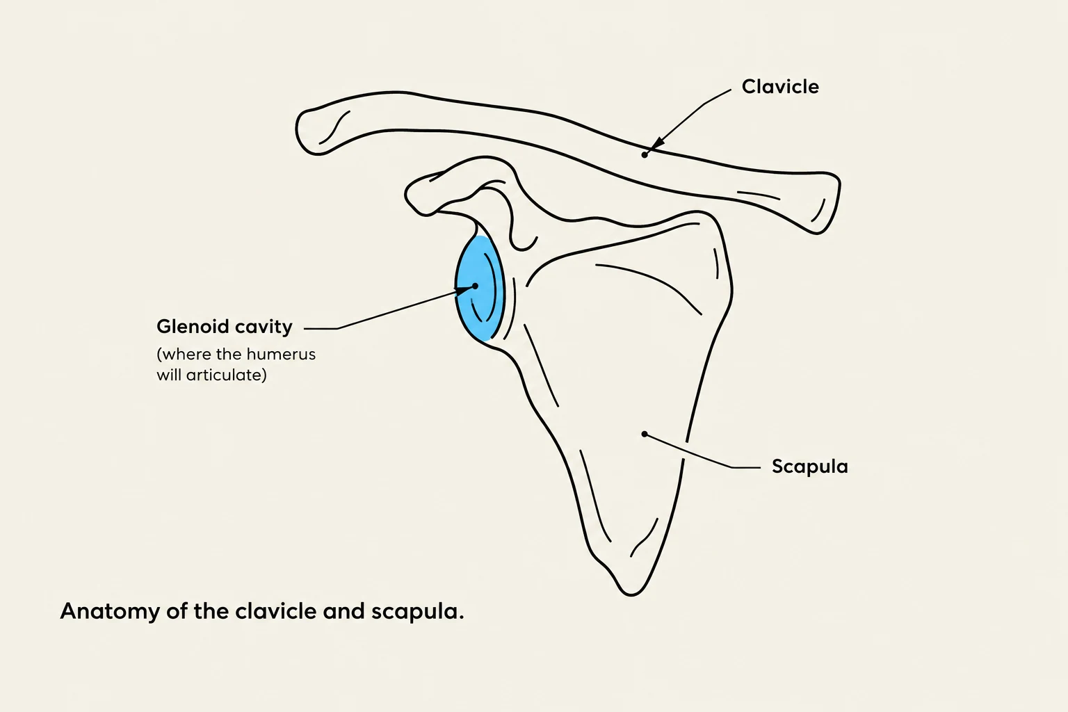
Anatomy of the clavicle and scapula.
The shoulder girdle, also known as the pectoral girdle, is the framework that connects the upper limbs to the axial skeleton. Unlike the pelvic girdle, which is a rigid ring designed for stability and weight-bearing, the shoulder girdle is designed for maximum mobility.
The girdle is primarily composed of two bones:
The Clavicle (Collarbone): Acts as a strut that holds the arm away from the torso, allowing for increased range of motion.
The Scapula (Shoulder Blade): A flat, triangular bone that sits on the posterior aspect of the thoracic cage.
The functional "joint" of the shoulder is the glenohumeral joint, where the head of the humerus meets the glenoid cavity of the scapula. Because the glenoid cavity is relatively shallow, the joint relies heavily on muscles, ligaments, and tendons (such as the rotator cuff) to maintain stability.
The Humerus
The humerus is the longest and largest bone of the upper limb. It is a long bone characterized by a proximal end, a shaft, and a distal end. The proximal portion is specifically adapted to articulate with the shoulder girdle to facilitate movement.
Proximal Humerus Anatomy
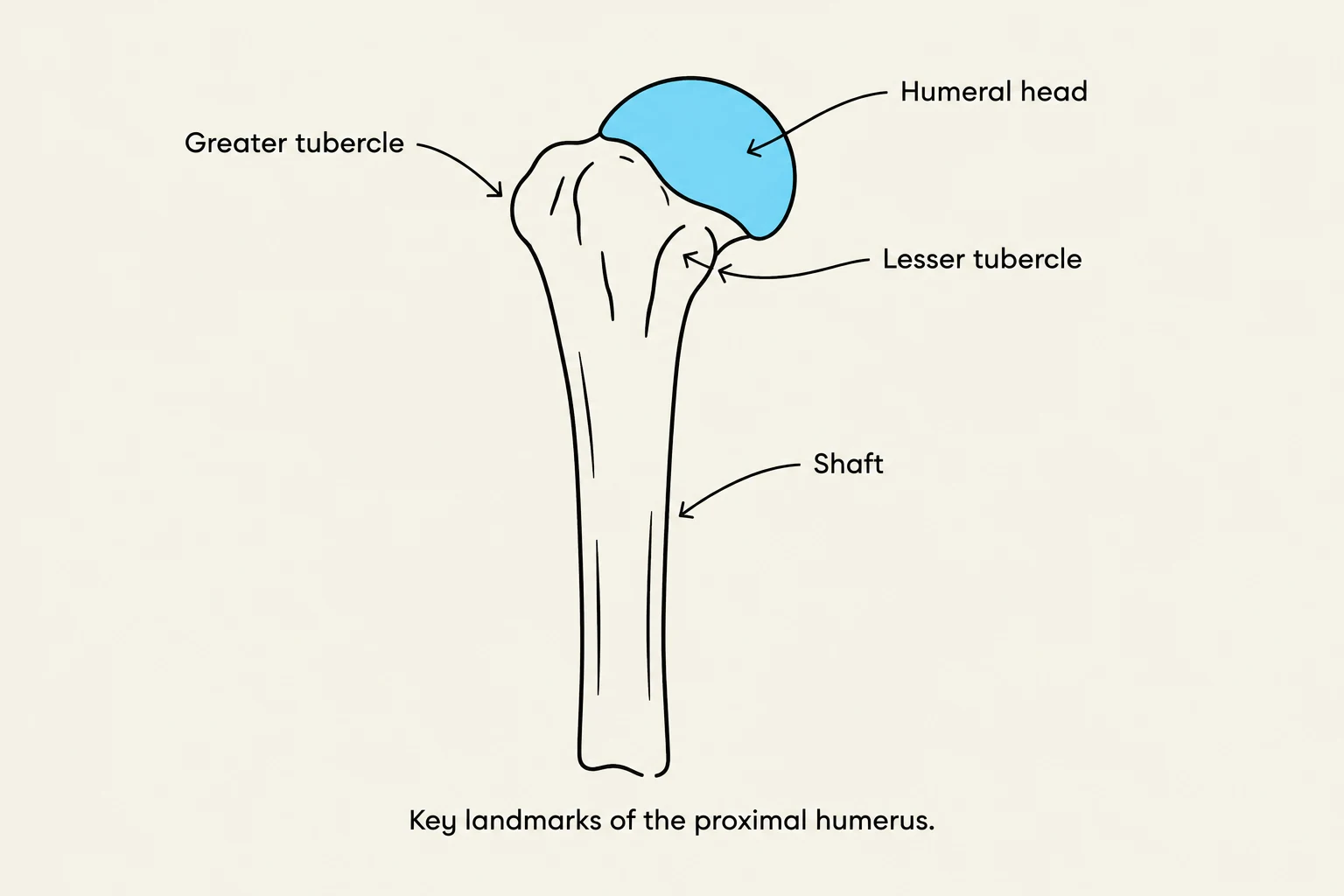
Key landmarks of the proximal humerus.
The proximal end of the humerus is the most critical area for shoulder function. Key anatomical landmarks include:
The Humeral Head: A smooth, rounded, hemispherical structure that fits into the glenoid cavity of the scapula.
The Shaft: The long, cylindrical body of the bone that extends distally toward the elbow.
The Greater and Lesser Tubercles: Bony prominences located near the head that serve as attachment points for the muscles of the rotator cuff.
In clinical practice, visualizing these structures clearly is essential for diagnosing fractures or dislocations. For instance, a lateral radiographic view is often used to assess the relationship between the humeral head and the glenoid cavity.
Fig. 3.10: A lateral view of the proximal humerus demonstrating the humeral head and its relationship to the surrounding thoracic structures.
Radiographic Evaluation of the Humerus
When performing imaging, such as a transthoracic lateral projection, specific criteria must be met to ensure a diagnostic image. A high-quality radiograph of the proximal humerus should demonstrate:
The Humeral Head: The rounded shape must be clearly visible to check for fractures or degenerative changes.
The Glenoid Cavity: The articulation point between the humerus and the scapula must be shown.
The Proximal Shaft: At least the proximal half of the humeral shaft should be clearly visualized.
Exam Tip: Breathing Techniques
When imaging the proximal humerus using a transthoracic lateral approach, clinicians often use a "breathing technique." This involves using a longer exposure time to blur overlying ribs and lung markings, which helps reduce visual interference and makes the bone structures easier to evaluate.
Summary Table: Key Anatomical Components
Structure
Component Type
Primary Function
Clavicle
Bone (Shoulder Girdle)
Provides structural support and spacing for the arm.
Scapula
Bone (Shoulder Girdle)
Provides the glenoid cavity for articulation.
Humeral Head
Proximal Humerus
Articulates with the scapula to allow rotation.
Humeral Shaft
Humerus Body
Provides leverage for muscle attachment and movement.
By understanding the relationship between the shoulder girdle and the humerus, healthcare professionals can better interpret imaging and understand the biomechanics of the human arm.
Keep reading for free.
Create an account to finish this reading and make it part of your study routine.