An overview of the human skull, detailing its two main divisions: the neurocranium (cranial skeleton) and the viscerocranium (facial skeleton). It describes the key bones within each division, including the frontal, parietal, temporal, ethmoid, sphenoid, maxillae, palatine, zygomatic, lacrimal, nasal, and mandible bone
The human skull is a complex assembly of bones that serves two primary functions: protecting the delicate tissues of the brain and providing the structural framework for the face, including the sensory organs and the mechanisms for mastication (chewing). To understand its intricate design, anatomists divide the skull into two main functional groups: the neurocranium and the viscerocranium.
The Two Divisions of the Skull
The skull is not a single solid unit but a collection of bones held together by specialized joints. These joints, known as sutures, are fibrous connections between the bones.
1. The Neurocranium (Cranial Skeleton)
The neurocranium is the portion of the skull that forms a protective "vault" or container for the brain. It consists of eight bones that enclose the cranial cavity.
2. The Viscerocranium (Facial Skeleton)
The viscerocranium comprises the bones that form the structure of the face. These bones provide the framework for the eyes, nose, and mouth, and they support the structures necessary for facial expression and feeding.
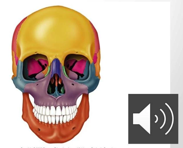
An illustration of the human skull, with different bones colored for clarity.
Detailed Anatomy of the Neurocranium
The neurocranium is composed of several bones that fit together like a puzzle to shield the central nervous system.
The Frontal Bone
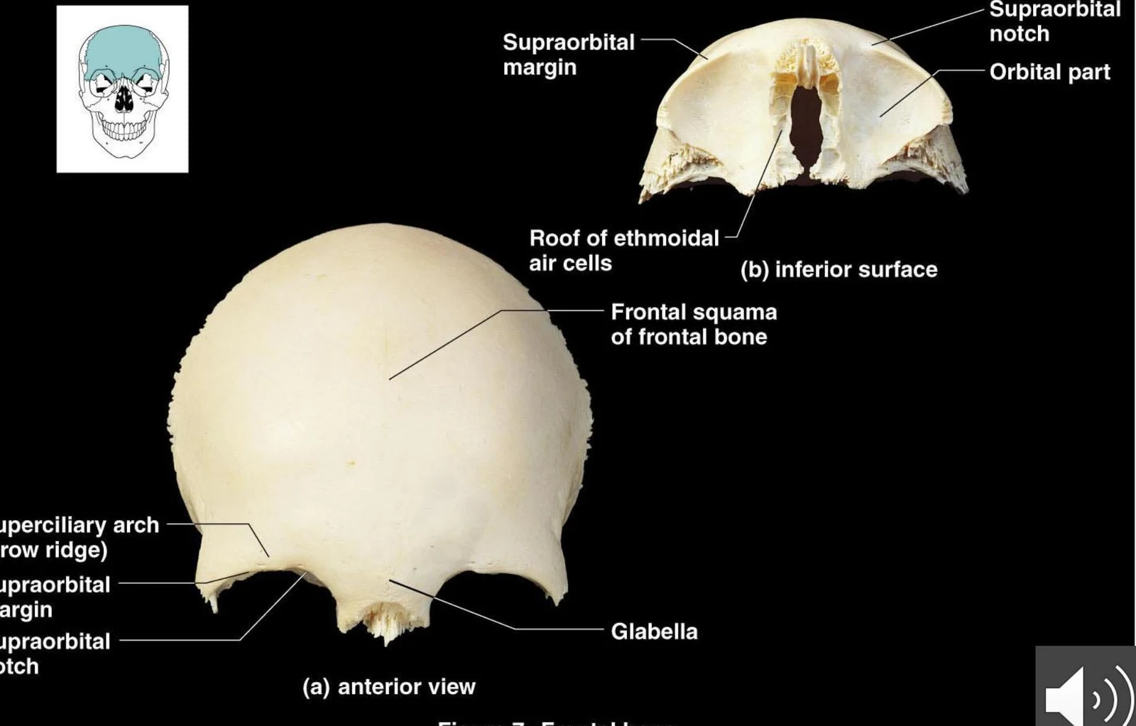
The frontal bone is a paired bone that, through the process of ossification during adulthood, forms a single unit. It is responsible for forming the bony forehead, the anterior portion of the brain case, and the roof of the orbital cavity (the eye socket).
Anatomical illustration of the frontal bone, showing anterior and inferior views with labels for key features such as the supraorbital margin, glabella, and frontal squama.Figure 7 Frontal bone.
The Parietal Bones
The parietal bones are large, paired bones that make up the majority of the superior (top) and lateral (side) surfaces of the cranium.
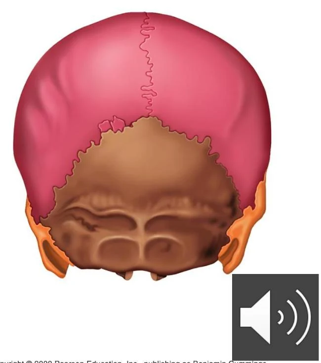
Posterior view of the human skull, highlighting the occipital bone (pink), parietal bones (brown), and temporal bones (orange).
The Temporal Bones
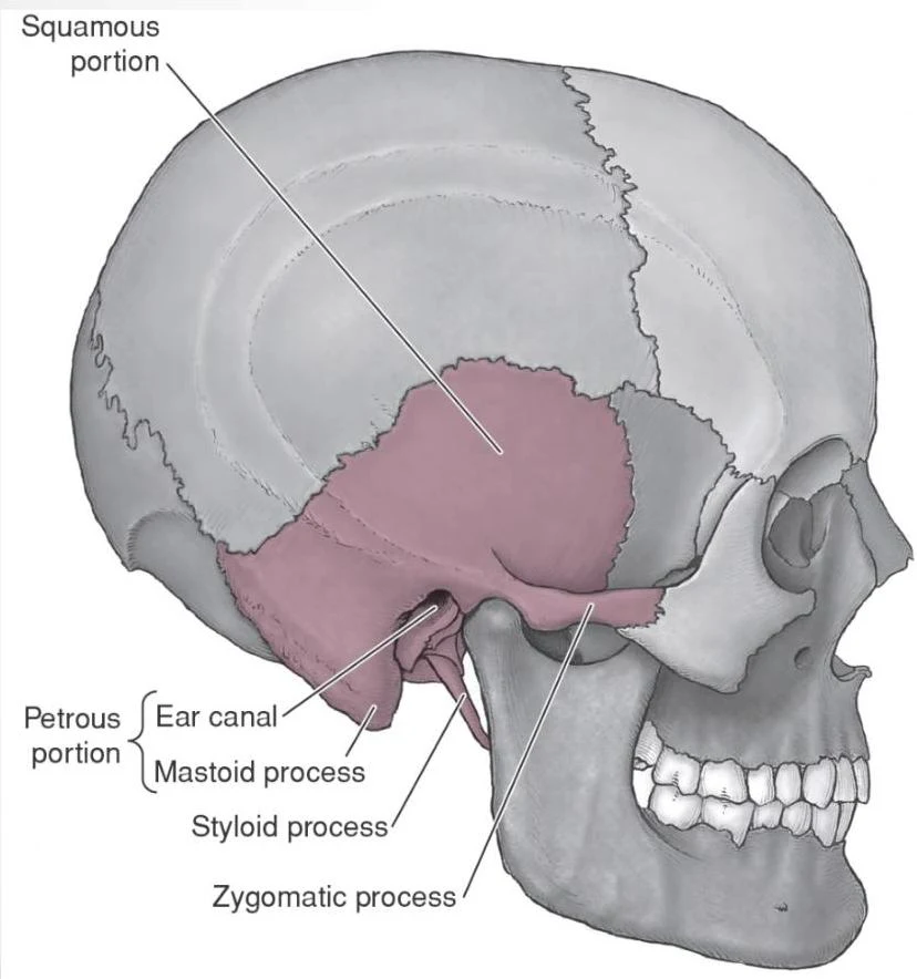
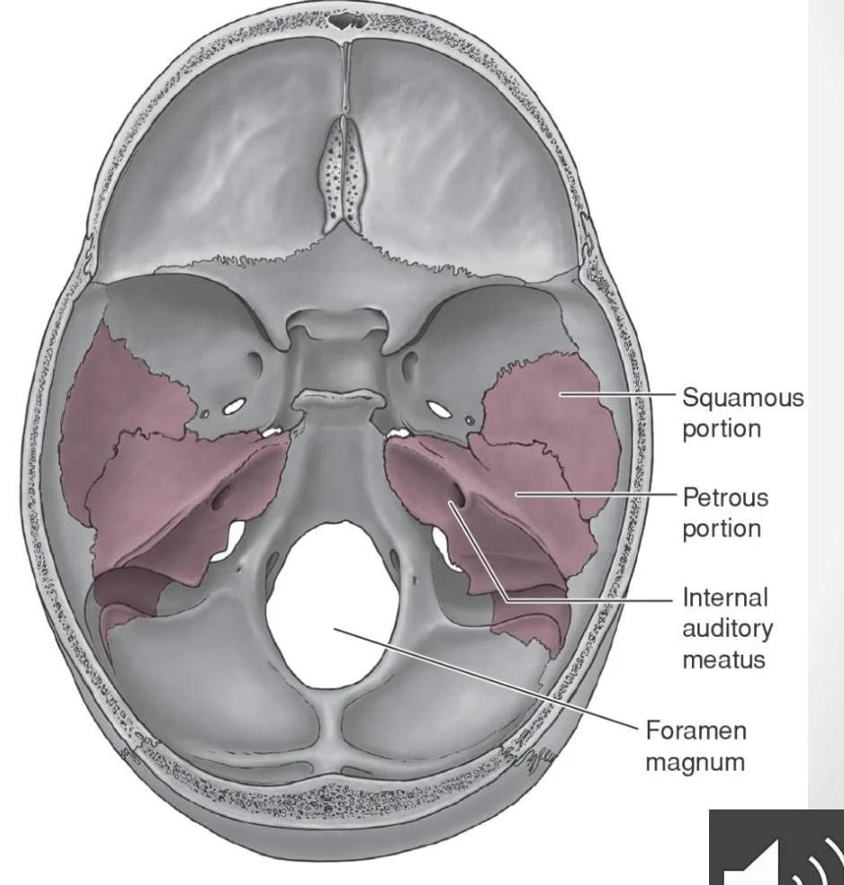
The temporal bones are paired structures that form the lateral base and the sides of the brain case. They are highly complex because they house structures related to hearing and balance. Key landmarks include the zygomatic process, the mandibular fossa (which forms part of the jaw joint), the mastoid process, the styloid process, and the external auditory meatus (the ear canal).
An illustration of the human skull, highlighting the squamous portion, petrous portion, ear canal, mastoid process, styloid process, and zygomatic process.
Anatomical illustration of the inferior view of the human skull, highlighting the squamous portion, petrous portion, internal auditory meatus, and foramen magnum.
The Ethmoid and Sphenoid Bones
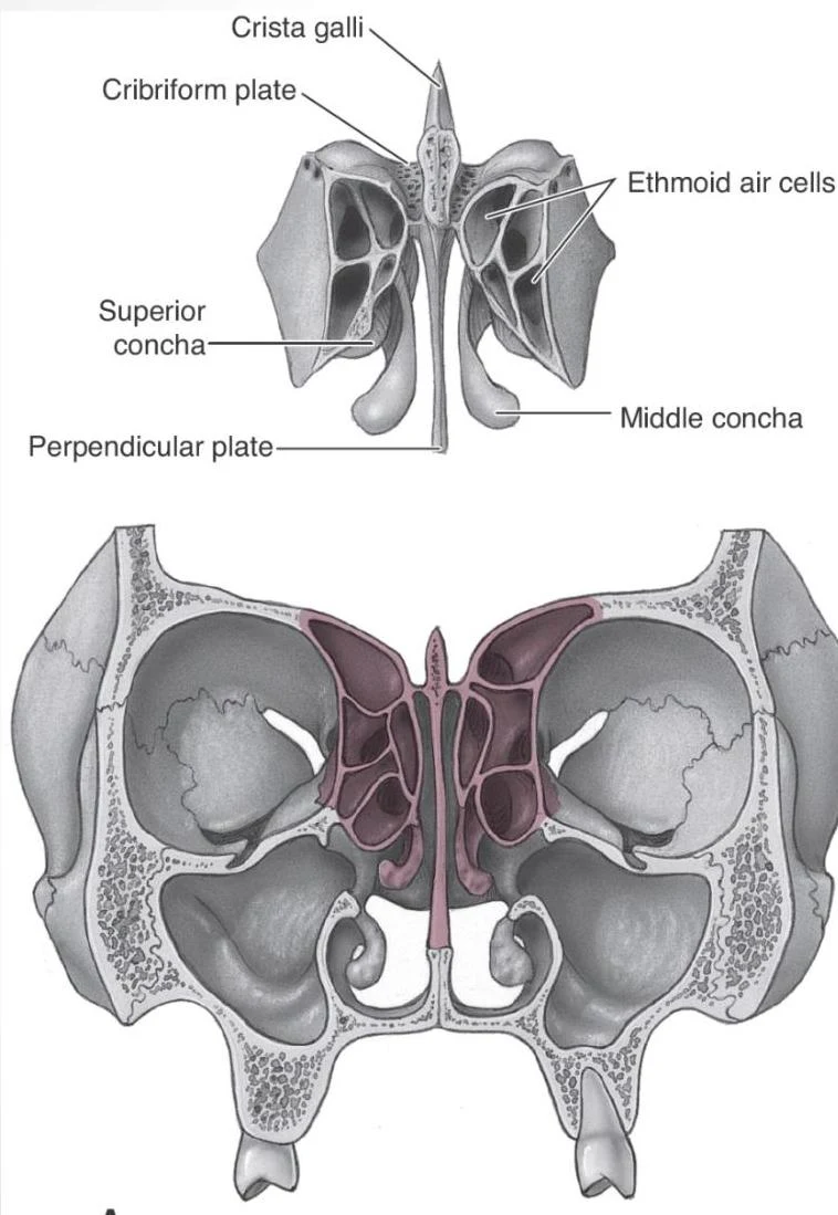
The ethmoid bone is a delicate, complex structure located at the core of the cranium. It contributes to the walls of the cranial, nasal, and orbital spaces. Notable features include the crista galli and the perpendicular plate.
Anatomical illustration of the ethmoid bone, showing the ethmoid air cells, crista galli, cribriform plate, superior concha, middle concha, and perpendicular plate.
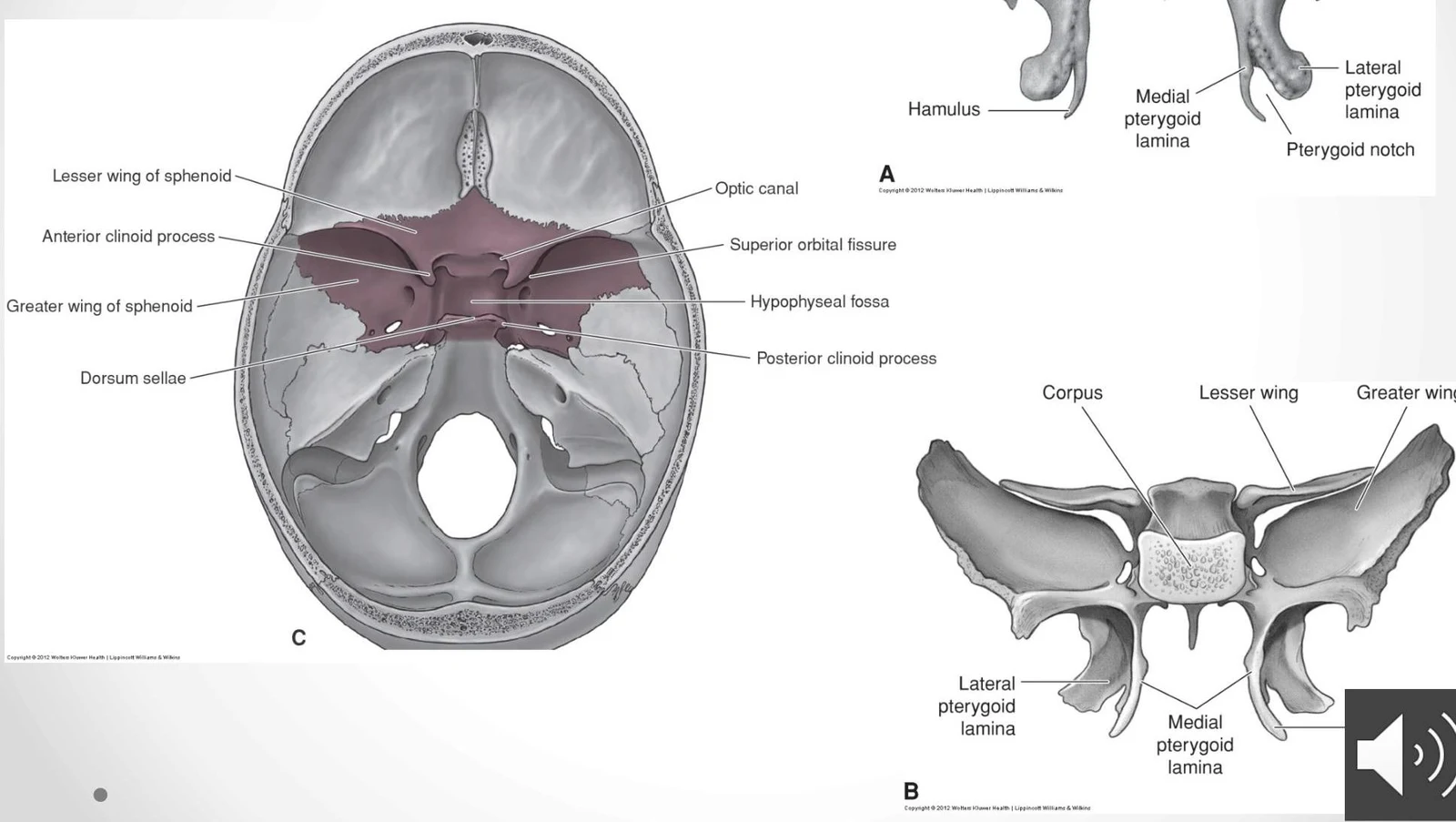
The sphenoid bone is arguably the most complex bone, situated at the base of the skull. It acts as a central structural hub. Its anatomy includes a central body (corpus), lesser wings located superior to the body, greater wings, and the medial and lateral pterygoid plates that project downward.
Anatomical illustration of the sphenoid bone, showing its various parts and their locations within the skull.
Study Tip: When identifying the sphenoid bone, look for the "Sella turcica," a saddle-shaped depression in the body of the bone that holds the pituitary gland.
Detailed Anatomy of the Viscerocranium
The viscerocranium forms the face and includes several bones that interact with the sensory organs and the jaw.
The Maxillae and Palatine Bones
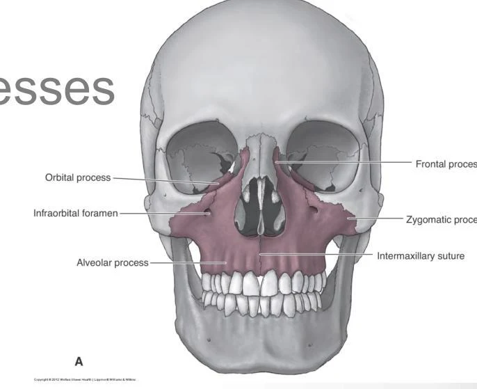
The maxillae are paired bones that form the upper dental arch, the roof of the mouth, and part of the nose. Each maxilla consists of a body and four distinct processes: the zygomatic, frontal, alveolar, and palatine processes.
An illustration of the human skull highlighting the maxilla, including labels for the orbital process, infraorbital foramen, alveolar process, frontal process, zygomatic process, and intermaxillary suture.
The palatine bones are also paired and contribute to the formation of the nasal, orbital, and oral cavities. They feature a horizontal process that helps form the hard palate.
Zygomatic and Lacrimal Bones
The zygomatic bones (cheekbones) are paired, quadrilateral-shaped bones that articulate with the maxilla, frontal, temporal, and sphenoid bones.
The lacrimal bones are the smallest bones of the face. They are paired and form part of the medial wall of the orbital cavity and the lateral wall of the nasal cavity.
Nasal Bones and the Orbital Cavity
The nasal bones are paired bones that form the bony bridge of the nose.
The orbital cavity (eye socket) is a complex space formed by the convergence of several bones. The roof is formed by the lesser wing of the sphenoid and the orbital plate of the frontal bone. The floor is composed of the orbital process of the palatine bone, the orbital surface of the maxillary bone, and the zygomatic bone.
Exam Note: The medial wall of the orbit is a composite structure involving the sphenoid body, the ethmoid bone, the frontal process of the maxilla, the lacrimal bone, and the nasal bone.
The Mandible and Nasal Septum
To complete the facial structure, we look at the bones responsible for the lower jaw and the division of the nasal passages.
The Mandible
The mandible is a single, horseshoe-shaped bone (which becomes a single bone by approximately age 2). It consists of a body and two vertical rami. A critical functional feature is the head of the condylar process, which articulates with the mandibular fossa of the temporal bone to form the temporomandibular (TMJ) joint.
The Nasal Septum
The nasal septum is the partition that divides the nasal cavity. It is formed by the vomer (which forms the inferior and posterior portion) and the perpendicular plate of the ethmoid bone.
Keep reading for free.
Create an account to finish this reading and make it part of your study routine.