


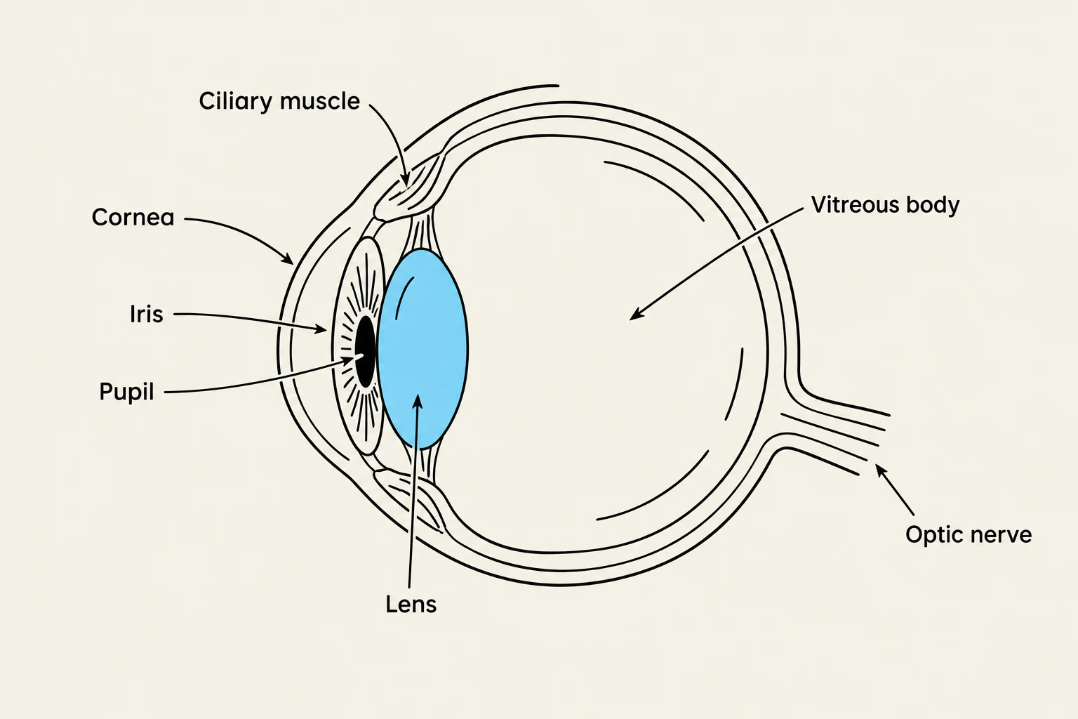
The human eye is a sophisticated sensory organ designed to capture light and convert it into electrical signals that the brain interprets as vision. To achieve this, the eye utilizes a complex arrangement of protective layers, refractive elements, and specialized neural tissues. Understanding its anatomy requires looking at three distinct perspectives: the external protective structures, the internal layers and chambers, and the supporting systems that manage movement and lubrication.
The External Protective Layers and Landmarks
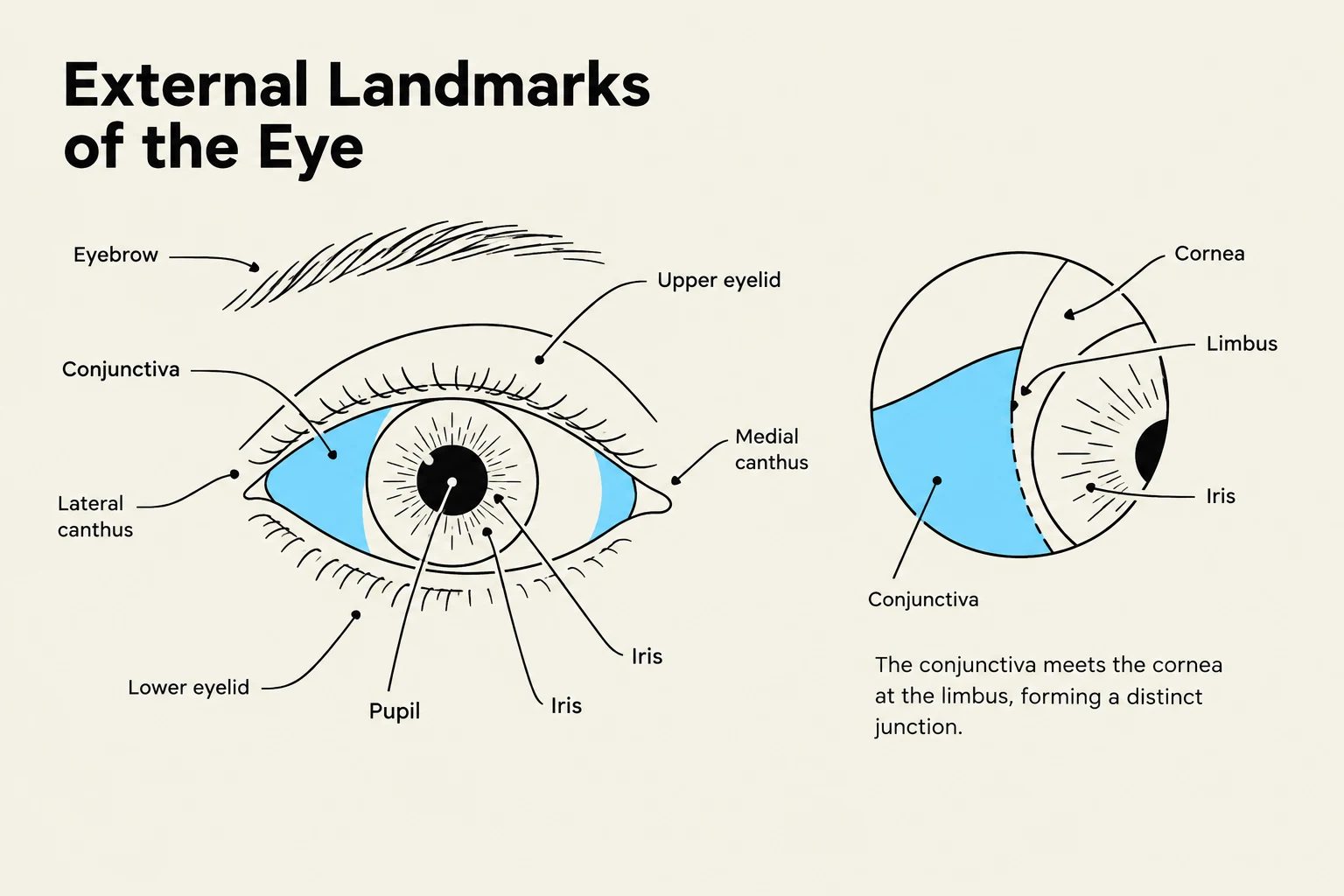
The eye is first protected by several layers of tissue that safeguard the delicate internal components. The outermost layer is the sclera, a dense, white, and avascular structure that provides physical support and maintains the eye's shape. Moving toward the front of the eye, the sclera transitions into the cornea, an optically clear structure that constitutes the anterior sixth of the globe. The specific junction where the transparent cornea meets the white sclera is known as the limbus.
Covering the front of the eye and the inside of the eyelids is the conjunctiva, a thin, clear mucous membrane. It is divided into two parts: the palpebral conjunctiva, which coats the eyelids, and the bulbar (or ocular) conjunctiva, which covers the outer surface of the eye (excluding the cornea).
Around the eye, landmarks such as the inner canthus (the medial corner) and the outer canthus (the lateral corner) define the boundaries of the ocular region. At the inner corner, one can often find the caruncle, a small, fleshy, pink nodule.
Keep reading for free.
Create an account to finish this reading and make it part of your study routine.
Continue reading freeAlready have an account? Log in
Updated