


The human hand is a masterpiece of biological engineering, providing a unique combination of strength and fine motor dexterity. At the base of the hand lies the wrist, a complex junction that facilitates a wide range of motion. This movement is made possible by a cluster of eight small, irregularly shaped bones known as the carpal bones. These bones act as a bridge, transitioning the structural support of the forearm to the long bones of the hand, known as the metacarpals, and eventually to the phalanges (finger bones).
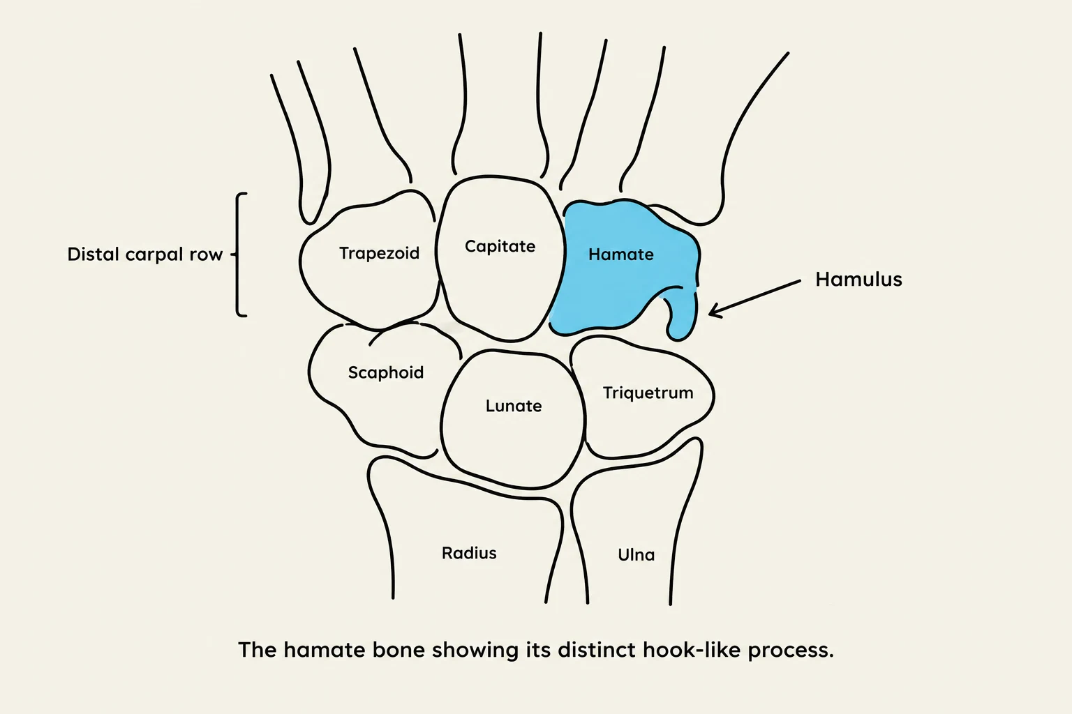
To understand the wrist, one must view the carpal bones not as a random pile of fragments, but as a highly organized system divided into two distinct rows: the proximal row and the distal row.
Figure 1: Anatomical line drawing of the human hand and wrist showing the color-coded carpal bones.
The Proximal Row: The Foundation of Wrist Motion

- Scaphoid: Located on the lateral side of the wrist, the scaphoid is a critical link between the forearm and the distal row. Due to its position and blood supply, it is one of the most frequently fractured bones in the wrist.
- Lunate: Situated in the center of the proximal row, the lunate acts as a central pivot point.
- Triquetrum: Positioned on the medial side of the proximal row, providing stability to the ulnar side of the wrist.
- Pisiform: This is a unique, small, pea-shaped bone that sits superficially on top of the triquetrum. Unlike the other carpal bones, it is a sesamoid bone, meaning it is embedded within a tendon.

Keep reading for free.
Create an account to finish this reading and make it part of your study routine.
Continue reading freeAlready have an account? Log in
Updated