Anatomy of the Human Brain and its Protective Structures
Explore the intricate anatomy of the human brain, including its gray and white matter, the diencephalon, brainstem, and the protective meninges. Learn about the ventricular system and the functions of cerebrospinal fluid (CSF).
The human brain is a complex organ composed of specialized tissues and protective structures designed to maintain homeostasis and process information. To understand how the brain functions, one must look beyond the surface and examine its internal cellular composition, the deep-seated regulatory centers, the vital brainstem, and the intricate systems that protect and nourish these delicate tissues.
Internal Composition: Gray and White Matter
The brain is not a uniform mass of tissue; rather, it is organized into two distinct types of matter based on the components of the neurons present.
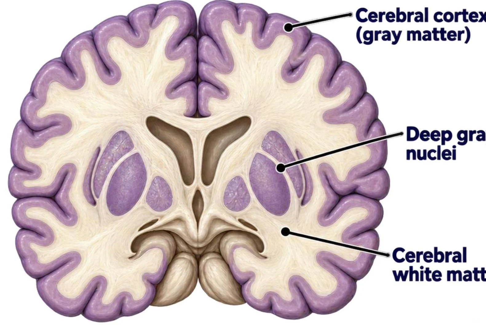
Gray matter consists of neuron cell bodies, dendrites, and synapses. In the cerebrum, gray matter is located superficially, forming the cerebral cortex. This area is where much of the high-level processing, such as sensation and cognition, occurs.
White matter consists primarily of myelinated axons that form tracts. These axons act as the "wiring" of the brain, allowing communication between different regions. In the brain, white matter is located deep to the cerebral cortex.
This is a cross-section of the human brain, illustrating the cerebral cortex (gray matter), deep gray nuclei, and cerebral white matter.
Study Tip: Remember that the arrangement of gray and white matter in the brain is the reverse of what you will find in the spinal cord. In the brain, gray matter is superficial; in the spinal cord, it is central.
The Diencephalon
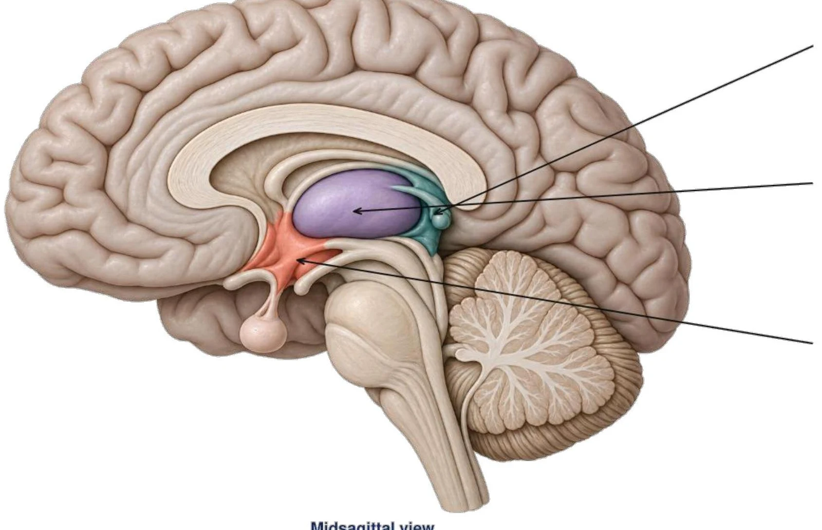
Located deep to the cerebrum and positioned between the cerebrum and the brainstem, the diencephalon serves as a central hub for sensory and autonomic integration. It surrounds the third ventricle. The diencephalon is composed of three major components:
Thalamus: This structure acts as a relay station, sending most sensory information to the cerebral cortex and assisting in motor processing.
Hypothalamus: Located inferior and anterior to the thalamus, it forms the floor of the third ventricle. It is critical for maintaining homeostasis by regulating autonomic and endocrine activity, including temperature, hunger, thirst, and circadian rhythms.
Epithalamus: This region includes the pineal gland, which secretes melatonin to help regulate circadian rhythms.
Midsagittal view of the human brain showing key structures.
The Brainstem
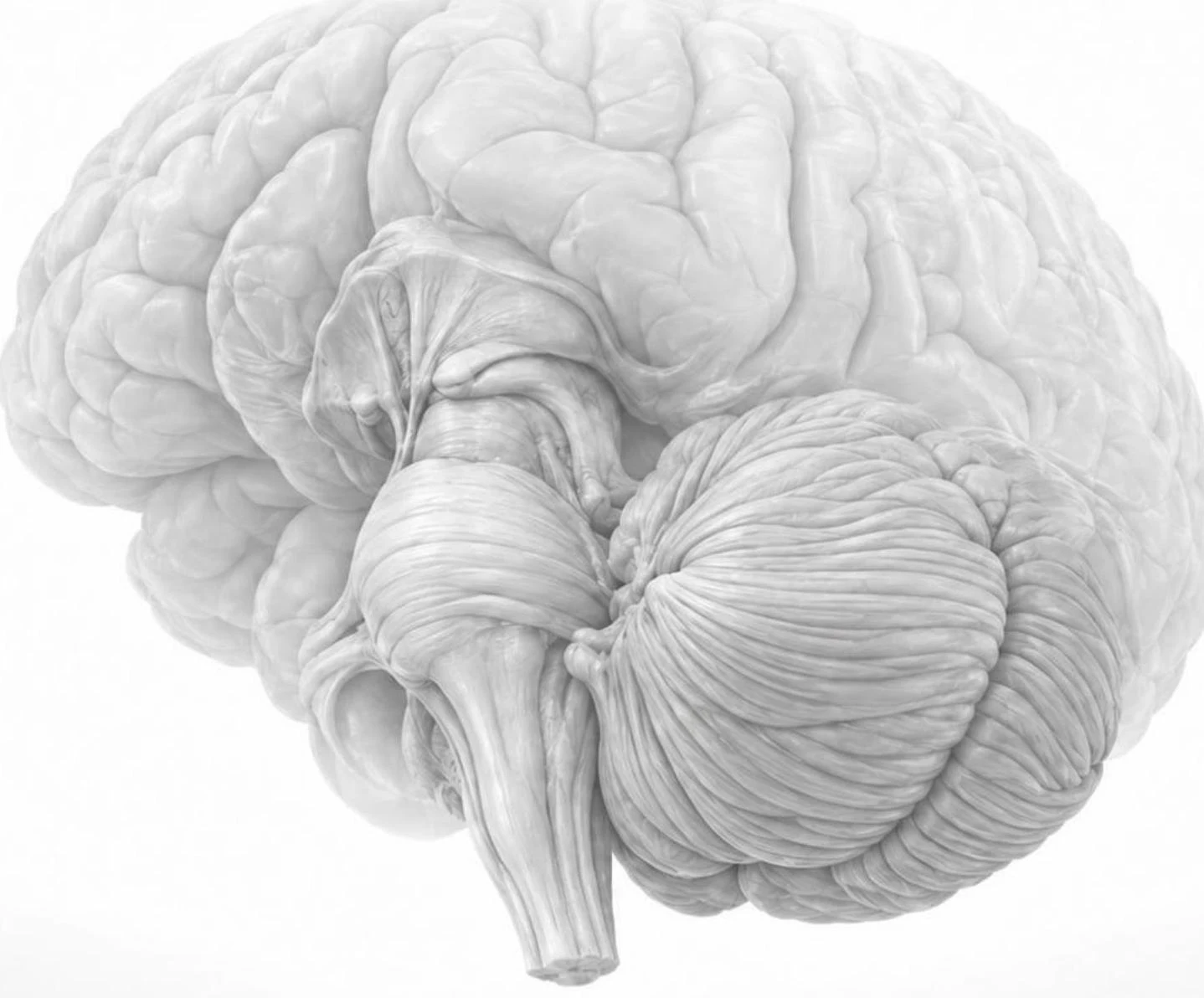
The brainstem is the vital connection between the brain and the spinal cord. It contains essential pathways and autonomic centers that control life-sustaining functions. It is divided into three main regions:
Midbrain
The midbrain (mesencephalon) is the most superior part of the brainstem. It coordinates visual and auditory orienting reflexes and contributes to motor control and alertness.
Pons
The pons acts as a bridge, relaying information between the cerebrum and the cerebellum. It also assists in respiratory control and supports functions like sleep, facial sensation, and chewing.
Medulla Oblongata
The medulla oblongata is the most inferior part of the brainstem, extending to the spinal cord at the foramen magnum. It contains vital autonomic centers that regulate heart rate, blood pressure, and breathing. It also coordinates reflexes such as swallowing, coughing, and vomiting.
A detailed illustration of the human brain, highlighting the cerebellum and brainstem.
Protection: The Meninges
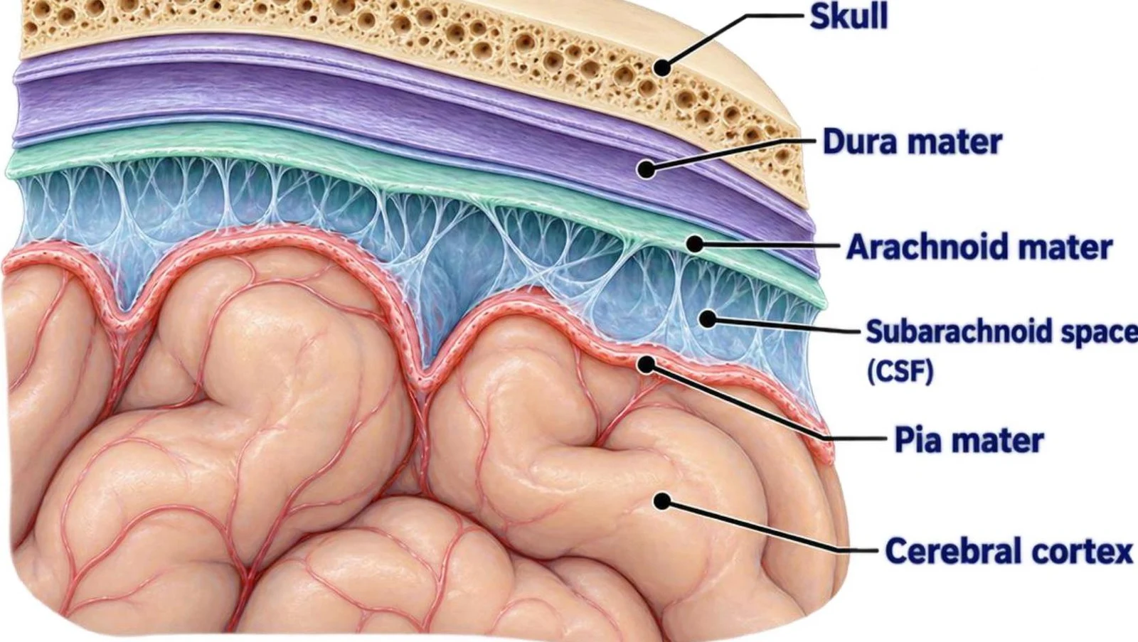
To safeguard the delicate neural tissue from mechanical injury, the brain is encased in the skull and wrapped in three protective layers of connective tissue known as the meninges.
Dura mater: The outermost layer, which is tough and durable.
Arachnoid mater: The middle layer, characterized by a web-like appearance.
Pia mater: The innermost layer, a delicate membrane that follows the contours of the brain's surface.
Diagram illustrating the layers of the meninges surrounding the brain, including the skull, dura mater, arachnoid mater, subarachnoid space containing cerebrospinal fluid (CSF), pia mater, and the cerebral cortex.
The Ventricular System and Cerebrospinal Fluid (CSF)
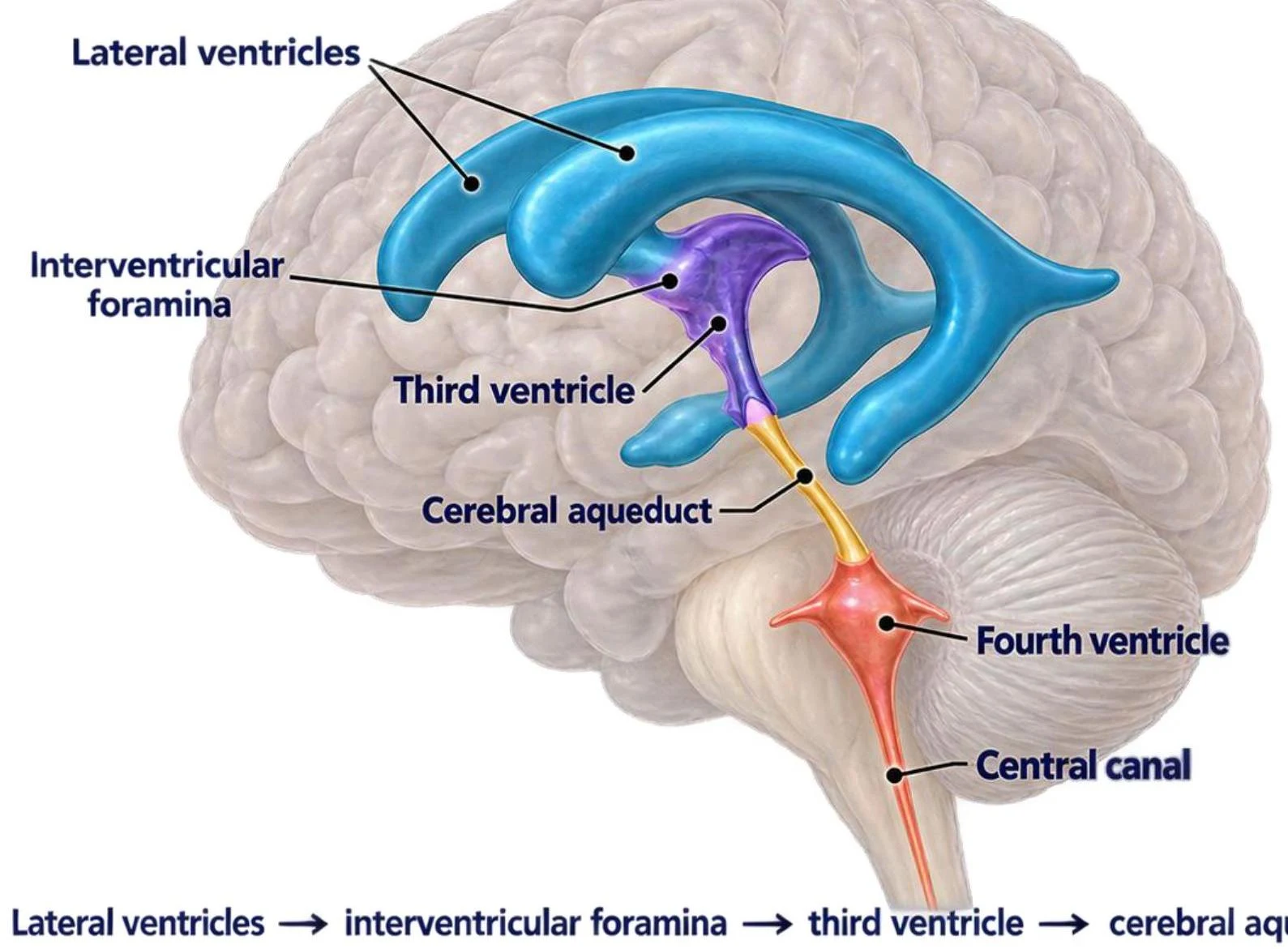
The brain contains a network of interconnected, fluid-filled chambers called ventricles. These chambers are lined with ependymal cells and are filled with cerebrospinal fluid (CSF).
Functions of CSF
CSF is continuously produced and returned to the venous bloodstream, providing several critical services:
Buoyancy: It reduces the effective weight of the brain.
Protection: It acts as a cushion for the brain and spinal cord.
Chemical Stability: It maintains a controlled chemical environment for neurons.
Waste Transport: It helps remove metabolic waste from the central nervous system.
The Flow of CSF
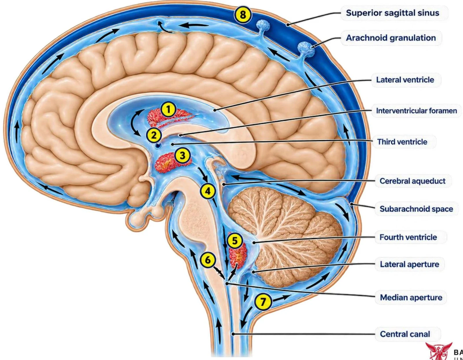
The movement of CSF through the brain follows a specific, regulated pathway:
Secretion: CSF is produced by the choroid plexuses within the lateral ventricles.
Interventricular Foramina: The fluid flows through these openings into the third ventricle.
Cerebral Aqueduct: From the third ventricle, CSF passes through this narrow channel into the fourth ventricle.
Apertures: The fluid exits the fourth ventricle through the median and lateral apertures.
Subarachnoid Space: Once outside the ventricles, the CSF enters the subarachnoid space, surrounding the brain and spinal cord.
Reabsorption: Finally, the CSF is returned to the dural venous sinuses via arachnoid granulations.
Diagram illustrating the ventricular system of the human brain, showing the lateral ventricles, interventricular foramina, third ventricle, cerebral aqueduct, fourth ventricle, and central canal.
Diagram illustrating the flow of cerebrospinal fluid through the ventricles and subarachnoid space of the brain.
Exam Tip: If asked about the production of CSF, always mention the choroid plexus. If asked about its return to the blood, mention the arachnoid granulations.
Keep reading for free.
Create an account to finish this reading and make it part of your study routine.