Detailed anatomical explanation of the human hip bone (coxal bone), femur, and patella, including their components, articulations, and mechanical functions in locomotion.
The human lower appendage is a sophisticated mechanical system designed for two primary purposes: supporting the weight of the entire body and facilitating locomotion. This structural integrity is provided by a chain of heavy-duty bones—the hip bone, the femur, and the patella—which work in concert through specific articulations to allow for complex movements like walking, running, and jumping.
The Right Hip Bone (Coxal Bone)
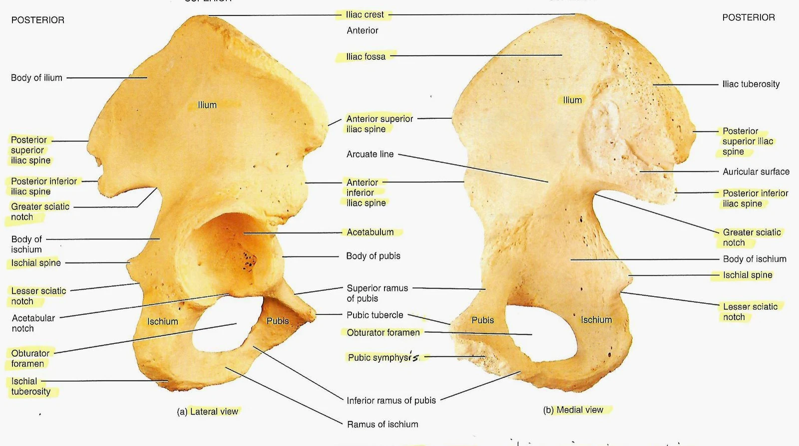
The foundation of the lower limb begins with the hip bone, also known as the coxal bone. This bone is not a single unit but is formed by the fusion of three distinct skeletal elements that meet at the acetabulum, the socket that receives the head of the femur.
Components of the Hip Bone
To understand the pelvic girdle, one must distinguish between its three primary parts:
Ilium: This is the most superior and largest portion of the hip bone. It forms the broad, flared upper region of the pelvis.
Ischium: Located in the posteroinferior position, the ischium forms the lower and back part of the hip bone.
Pubis: This is the anterior portion of the hip bone, forming the front of the pelvic structure.
Anatomical illustration of the human pelvis, showing lateral and medial views with labels for various bones and landmarks including the ilium, ischium, pubis, acetabulum, and obturator foramen.Figure 1.31 Right hip bone showing the ilium, ischium, and pubis.
Exam Tip: When identifying the hip bone in a lateral view, look for the large, wing-like ilium at the top and the ischium forming the base of the "seat" area.
The Right Femur
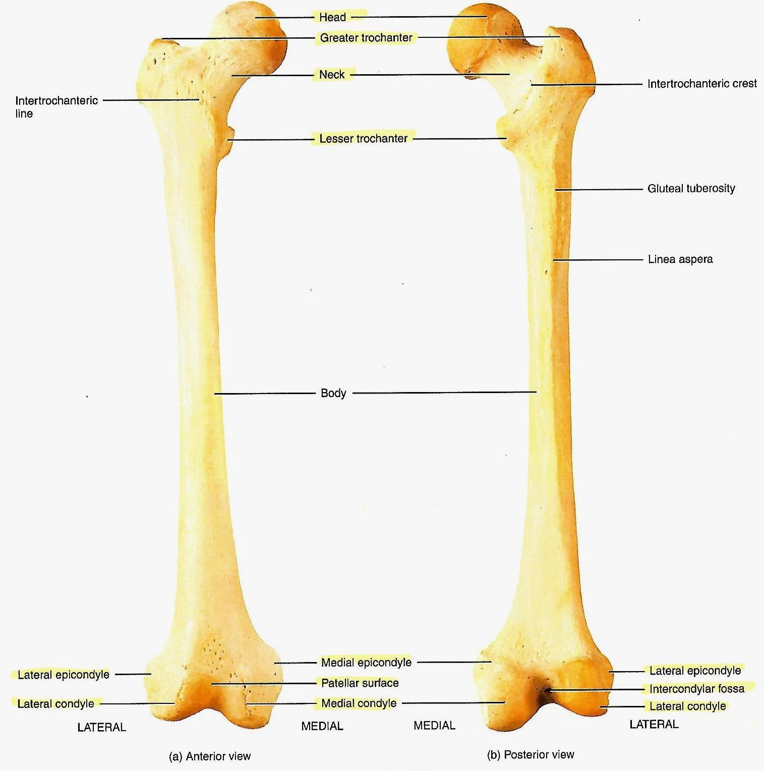
Moving distally from the pelvis, we encounter the femur, which is the longest, heaviest, and strongest bone in the human body. It serves as the primary structural element of the thigh.
Anatomical illustration of the anterior and posterior views of the human femur, labeling key features such as the head, neck, trochanters, body, and condyles.Figure 1.33 Right femur.
The Proximal End of the Femur
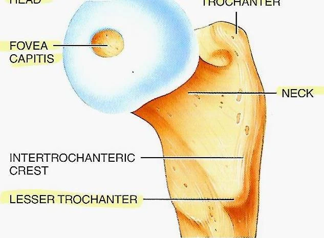
The proximal end (the top part) is the most critical area for hip articulation. It contains several landmarks that dictate how the leg moves and how muscles attach to the bone:
Head of the Femur: A rounded projection that fits into the acetabulum of the hip bone to create the hip joint.
Neck of the Femur: A constricted region that connects the head to the main shaft of the bone. This area is clinically significant due to its susceptibility to fractures.
Greater and Lesser Trochanters: These are large, bony prominences that serve as major attachment sites for the powerful muscles of the hip and thigh.
An illustration of the proximal femur, highlighting anatomical landmarks such as the fovea capitis, neck, intertrochanteric crest, and lesser trochanter.Figure 1.33 (c) Medial view of the proximal end of the femur.
The Distal End and Articulation
As the femur travels toward the knee, it widens at the distal end. This region is characterized by the medial and lateral condyles. These rounded projections are essential for the articulation of the knee joint, providing the surface area necessary for smooth movement when interacting with the tibia and the patella.
The Right Patella
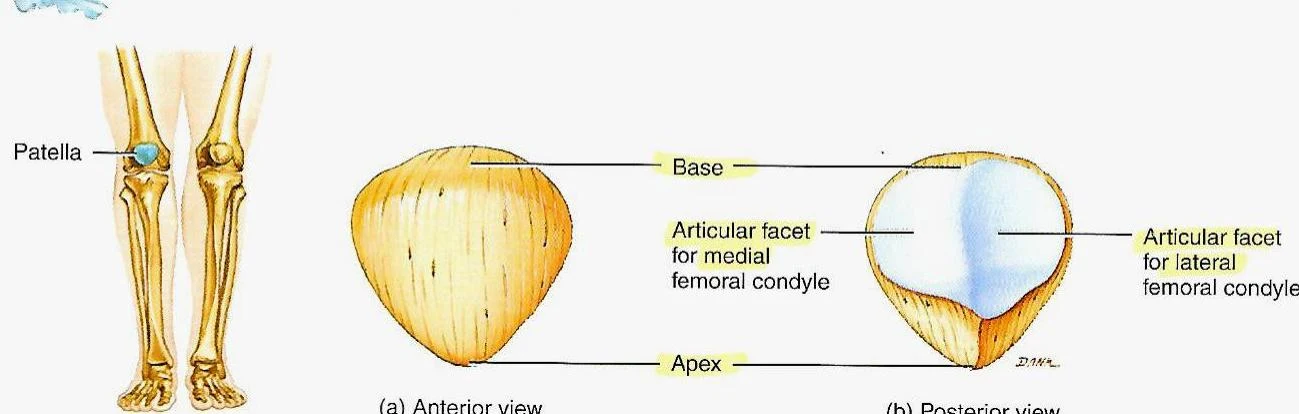
The patella, commonly referred to as the kneecap, is a unique type of bone known as a sesamoid bone. Sesamoid bones are embedded within tendons; the patella is specifically located anterior to the knee joint.
Anatomical illustration of the patella, showing anterior and posterior views with labels for its base, apex, and articular facets for the medial and lateral femoral condyles.Figure 8.14 Right patella.
Mechanical Function
The primary role of the patella is to act as a fulcrum. It articulates directly with the lateral and medial condyles of the femur. As the knee undergoes flexion (bending) and extension (straightening), the patella glides within the groove formed by these condyles. This movement provides a mechanical advantage to the quadriceps muscle group, allowing them to extend the leg more efficiently.
Summary of Structural Connections
To visualize the "structural chain" mentioned in the study materials, consider the following sequence of articulations:
Hip Joint: Formed by the articulation of the femoral head and the acetabulum of the hip bone (composed of the ilium, ischium, and pubis).
Knee Joint: Formed by the articulation of the femoral condyles (medial and lateral) with the patella and the upper part of the tibia.
Together, these bones create a stable yet mobile framework capable of managing the high mechanical loads encountered during human locomotion.
Clinical Note: Because the femur and hip bone bear the majority of body weight, injuries to the femoral neck or the acetabulum can significantly impact a person's ability to walk and maintain balance.
Keep reading for free.
Create an account to finish this reading and make it part of your study routine.