


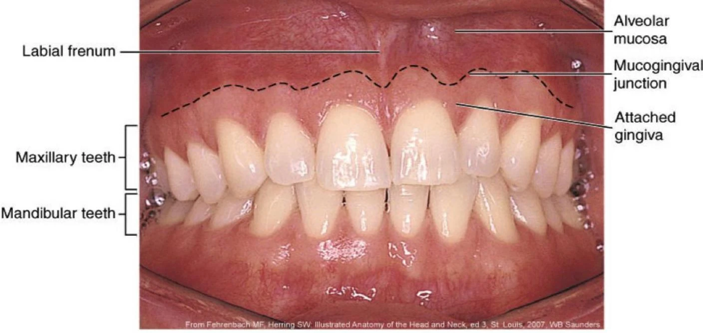
The oral cavity is a complex environment lined with specialized tissues designed to withstand the mechanical stresses of mastication (chewing) and the chemical challenges of the oral environment. To understand dental health and periodontal disease, one must first master the anatomy of the gingiva and the various types of oral mucosa. The gingiva is the specialized mucosa surrounding erupted teeth and represents the only visible portion of the periodontium.
The Gingival Complex: Free and Attached Gingiva
The gingiva is not a single uniform band of tissue; rather, it is composed of distinct zones that differ in their attachment to the tooth and their histological properties.
Marginal Gingiva and the Free Gingiva
The marginal gingiva (also known as the gingival margin) is the edge of the gingiva that is closest to the incisal edge of anterior teeth or the occlusal surface of posterior teeth. It acts like a "collar" around the tooth.
The free gingiva refers to the portion of the gingiva that is not tightly adhered to the underlying tooth surface. It surrounds the cervical enamel junction (CEJ) and extends to the base of the sulcus.
The Gingival Sulcus
The sulcus is the shallow space or "pocket" located between the tooth surface and the unattached cervical collar of the free gingiva. In a healthy mouth, this space is lined with nonkeratinized epithelium, and a clinician can measure its depth using a periodontal probe.
Clinical Note: A healthy sulcus depth is typically less than . If the depth increases, it may indicate the presence of a periodontal pocket, often resulting from bacterial activity and subsequent bone loss.
Keep reading for free.
Create an account to finish this reading and make it part of your study routine.
Continue reading freeAlready have an account? Log in
Updated