Anatomy of the Foot, Ankle, and Leg Muscles, Joints, and Retinacula
Explore the anatomy of the posterior compartment of the leg, the specialized joints of the foot and ankle, and the role of retinacula in maintaining structural integrity and facilitating movement.
The lower limb is a complex mechanical system designed to support body weight, facilitate locomotion, and provide stability during various activities. To understand how we walk, run, and balance, we must examine the intricate relationship between the muscles of the leg, the joints of the foot and ankle, and the connective tissues that guide them. This article explores the anatomy of the posterior compartment of the leg, the specialized joints of the foot, and the essential role of retinacula in maintaining structural integrity.
The Posterior Compartment of the Leg
The posterior compartment of the leg contains muscles that are primarily responsible for moving the foot and ankle. These muscles are organized into two distinct layers: the superficial group and the deep group. All muscles in this compartment share a common innervation: the tibial nerve.
Superficial Muscles
The superficial muscles are the most visible and powerful, providing the force necessary for movements like jumping or running.
Gastrocnemius: This muscle consists of two heads (medial and lateral) that originate from the postero-medial and postero-lateral femoral condyles, respectively. They insert into the posterior calcaneus via the powerful calcaneal (Achilles) tendon. Its primary function is ankle plantar flexion, and it also assists in knee flexion.
Soleus: Originating from the soleal line of the tibia and the posterior head/neck/shaft of the tibia, the soleus also inserts into the posterior calcaneus via the Achilles tendon. Like the gastrocnemius, its primary function is ankle plantar flexion.
Deep Muscles
Located beneath the superficial layer, the deep muscles provide more fine-tuned control over the toes and the stability of the foot.
Flexor Hallucis Longus (FHL): Originates from the posterior surface of the fibula and inserts into the plantar surface of the hallux (big toe). It is responsible for ankle plantar flexion and flexion of the big toe.
Flexor Digitorum Longus (FDL): Originates from the posterior surface of the tibia and inserts into the plantar surface of digits II–V. It facilitates ankle plantar flexion and flexion of the lesser toes.
Tibialis Posterior: This muscle originates from the posterior surfaces of both the tibia and fibula. It inserts into the plantar surface of nearly all tarsal and metatarsal bones. Its functions include ankle plantar flexion and inversion.
Diagram illustrating the superficial and deep layers of the calf muscles, including the gastrocnemius and soleus, and their relationship to the Achilles tendon and calcaneus.
Joint Mechanics and Movement
The movement of the foot and ankle is not the result of a single joint, but rather a coordinated effort across several articulations.
Ankle and Foot Movements
The most common movements associated with the ankle are plantar flexion (pointing the toes downward) and dorsiflexion (pulling the toes upward).
Beyond the sagittal plane, the foot can move in the frontal plane through inversion (turning the sole toward the midline) and eversion (turning the sole away from the midline). It is important to note that eversion and inversion of the foot occur at the subtalar joint, rather than the talocrural (ankle) joint.
Foot Articulations
The foot contains several types of joints that allow for different ranges of motion:
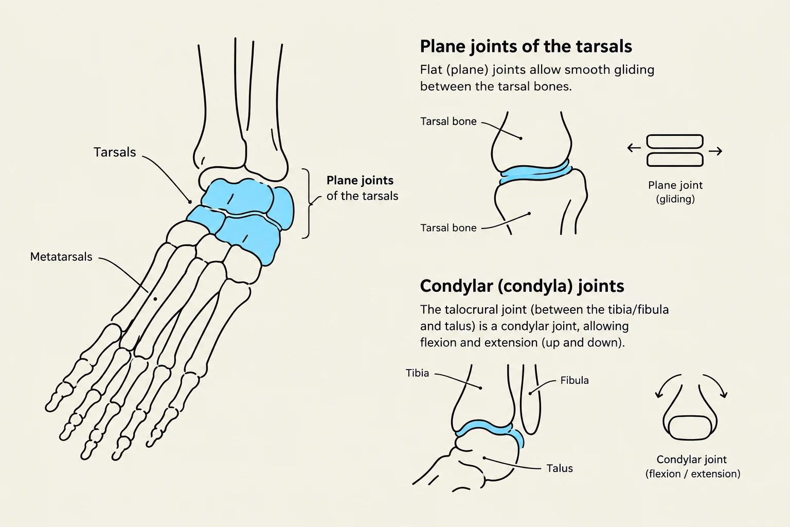
Intertarsal and Tarsometatarsal Joints: These are classified as plane joints, allowing for sliding or gliding movements.
Metatarsophalangeal (MTP) Joints: These are condylar joints that allow for flexion, extension, abduction, and adduction.
Interphalangeal Joints: These are hinge joints, providing flexion and extension of the toes.
Exam Tip: Abduction vs. Adduction of the Toes
When determining the movement of the toes, use Digit II as your reference point. Movement away from Digit II is abduction, while movement toward Digit II is adduction.
Diagrams illustrating plane joints of the tarsals and condylar joints of the ankle.
Retinacula: The "Pulleys" of the Leg
To ensure that muscles function efficiently, their tendons must be held close to the bone. This is the role of the retinacula—bands of thickened fascia that act like the eyelets or guides on a fishing rod.
If a retinaculum is injured, the tendon may no longer be held flush against the bone, causing it to "bowstring" and creating a straight line between its origin and insertion rather than following the contour of the joint.
The Five Primary Retinacula
There are five major retinacula located at the ankle and foot:
Superior Extensor Retinaculum: Holds down tendons of the anterior compartment.
Inferior Extensor Retinaculum: Also supports the anterior compartment.
Superior Fibular Retinaculum: Holds down tendons of the lateral compartment.
Inferior Fibular Retinaculum: Also supports the lateral compartment.
Flexor Retinaculum: Located on the medial aspect of the ankle.
The Tarsal Tunnel
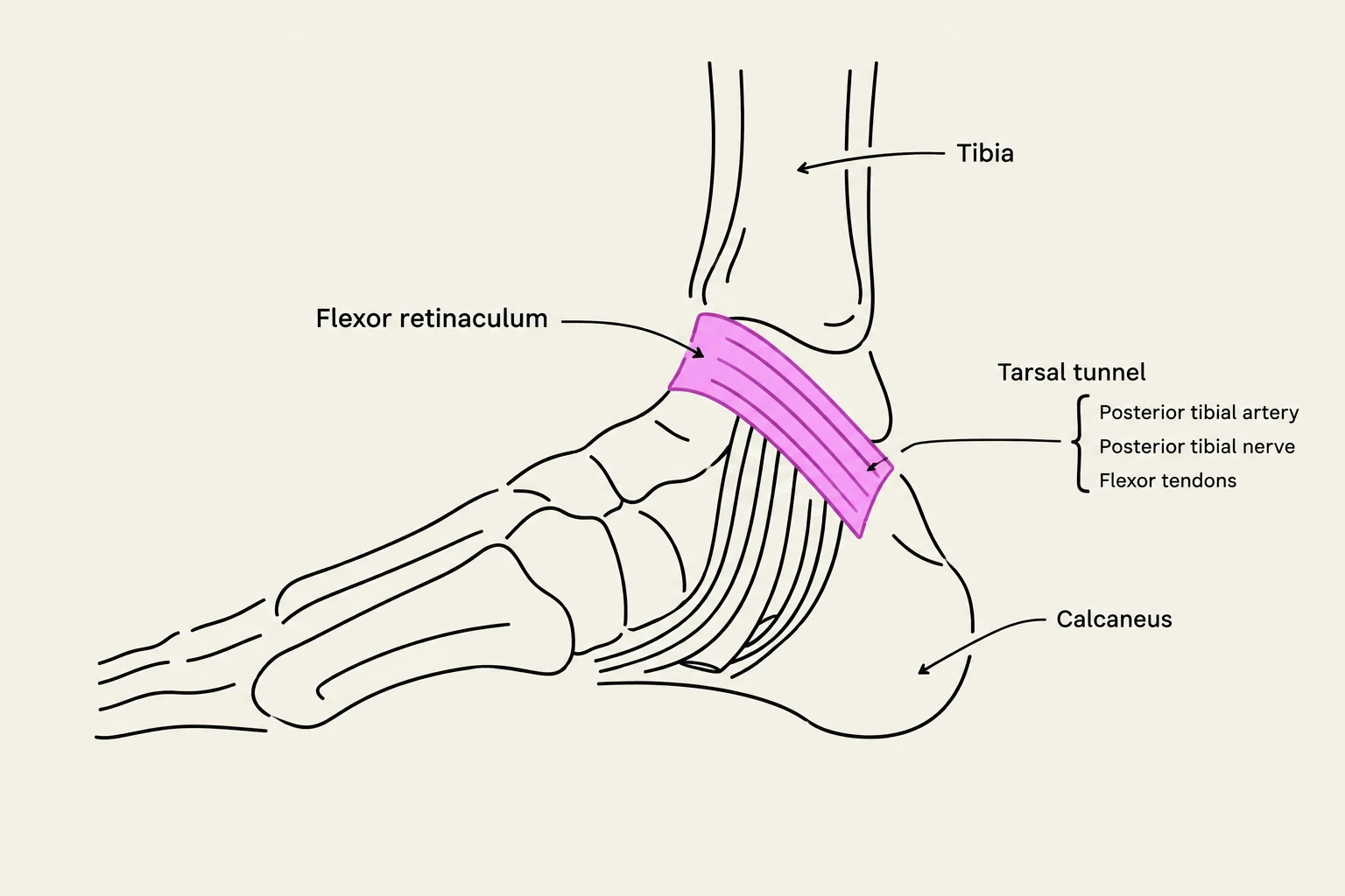
The flexor retinaculum plays a critical role in forming the tarsal tunnel, a hollow space on the posteromedial aspect of the ankle. This tunnel supports several vital structures, including tendons, nerves, and blood vessels.
To remember the order of structures passing through the tarsal tunnel from anterior to posterior, students often use the mnemonic: "Tom, Dick, And Very Nervous Harry":
Tibialis posterior tendon
Digitorum longus (Flexor) tendon
Artery (Tibial)
Vein (Tibial)
Nerve (Tibial)
Hallucis longus (Flexor) tendon
Anatomical illustration of the tarsal tunnel, showing the tibia, calcaneus, flexor retinaculum, and structures within the tunnel: posterior tibial artery, posterior tibial nerve, and flexor tendons.
Keep reading for free.
Create an account to finish this reading and make it part of your study routine.