An anatomical overview of the brachium (upper arm) and forearm, detailing their osteology, musculature, and innervation. Covers muscles responsible for flexion, extension, and pronation/supination, along with key bony landmarks and nerve pathways.
The upper limb is a complex mechanical system designed for both power and precision. To understand how we lift, rotate, and manipulate objects, we must examine the anatomical organization of the brachium (upper arm) and the forearm. These regions are divided into functional compartments—anterior, posterior, and lateral—defined by the muscles they contain, the nerves that supply them, and the bony structures that provide their framework.
Osteology: The Bony Framework
The movement of the arm and forearm is predicated on the structural integrity of the humerus, radius, and ulna.
The Distal Humerus
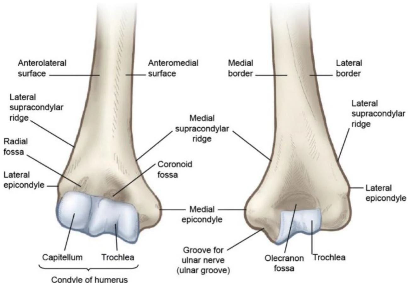
The distal humerus serves as the hinge for the elbow joint. Key landmarks include the medial and lateral epicondyles, which serve as the origins for the common flexor and extensor tendons, respectively. The trochlea and capitulum form the articular surfaces for the ulna and radius, while the olecranon and coronoid fossae accommodate the bony processes of the ulna during movement.
An illustration showing the anterior and posterior views of the distal humerus, labeling various anatomical landmarks such as the capitellum, trochlea, epicondyles, and fossae.
The Radius and Ulna
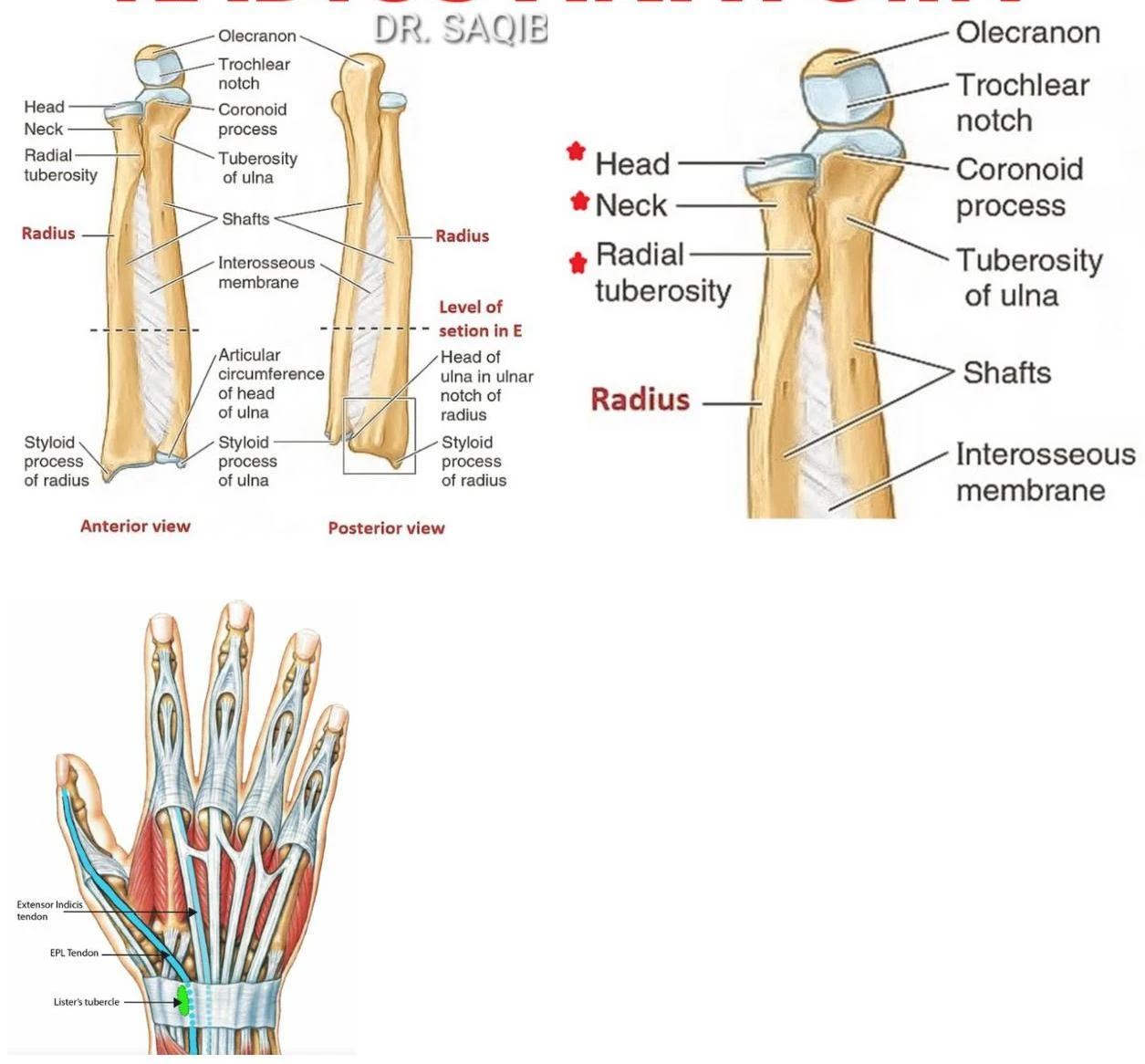
The forearm consists of two parallel bones: the ulna and the radius. The ulna is the primary stabilizer of the elbow, featuring the olecranon process (the "point" of the elbow) and the coronoid process. The radius is positioned laterally and is the primary bone involved in forearm rotation (pronation and supination). It provides the attachment point for the biceps brachii at the radial tuberosity.
Anatomical illustration of the human radius and ulna bones, showing anterior and posterior views, along with a detailed view of the tendons and structures in the hand.
Anatomical illustration of the ulna and radius bones of the human forearm, showing anterior and posterior views, and a detailed view of the ulna.
The Brachium (Upper Arm)
The brachium is divided into anterior and posterior compartments, primarily responsible for elbow flexion and extension.
Anterior Compartment (Flexors)
The muscles of the anterior brachium are primarily innervated by the musculocutaneous nerve ($C5, C6$):
Biceps Brachii: Attaches to the scapula (coracoid process and supraglenoid tubercle) and the radial tuberosity. It facilitates shoulder/elbow flexion and forearm supination.
Brachialis: Attaches to the anterior distal humerus and the coronoid process of the ulna; it is a pure elbow flexor.
Coracobrachialis: Originates at the coracoid process and assists in humeral flexion.
Posterior Compartment (Extensors)
The posterior compartment is dominated by the triceps brachii, which is innervated by the radial nerve ($C7, C8$):
Triceps Brachii: Features three heads (long, lateral, and medial) attaching to the scapula and humerus, with a distal attachment at the olecranon process of the ulna. It is the primary extensor of the elbow.
Exam Tip: Remember that the radial nerve supplies the entire posterior compartment of the arm and forearm. If a patient has "wrist drop," suspect radial nerve involvement.
The Forearm: Anterior-Medial Compartment
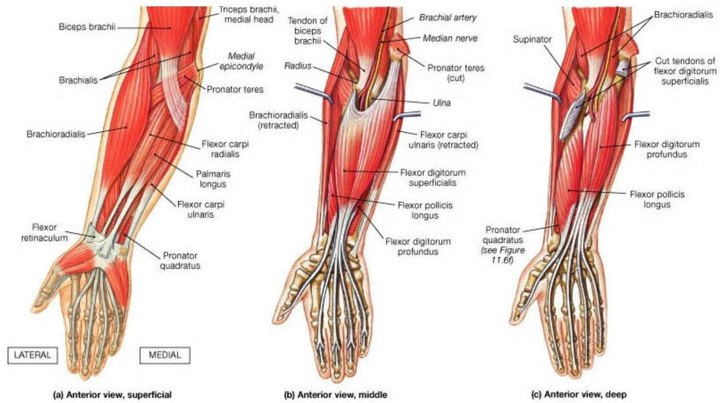
The anterior compartment of the forearm is primarily composed of muscles that flex the wrist and fingers or pronate the forearm. Most of these muscles originate from the medial humeral epicondyle via the common flexor tendon.
Superficial and Intermediate Layers
Pronator teres: Facilitates forearm pronation.
Flexor carpi radialis: Responsible for wrist flexion and radial deviation.
Palmaris longus: Assists in wrist flexion and tensions the palmar aponeurosis.
Flexor carpi ulnaris: Facilitates wrist flexion and ulnar deviation; notably, this muscle is innervated by the ulnar nerve.
Flexor digitorum superficialis: An intermediate muscle that flexes the MCP and PIP joints of digits 2–5.
Deep Layer
Flexor digitorum profundus: Flexes the MCP, PIP, and DIP joints. Its innervation is split: the medial half (digits 4–5) is supplied by the ulnar nerve, while the lateral half (digits 2–3) is supplied by the anterior interosseous nerve.
Flexor pollicis longus: Manages flexion of the thumb; innervated by the anterior interosseous nerve.
Pronator quadratus: A deep muscle that assists in forearm pronation.
Most of these muscles are innervated by the median nerve, with the exceptions noted above.
Anatomical illustration of the anterior view of the muscles and bones of the human forearm, showing superficial, middle, and deep layers.
The Forearm: Posterior-Lateral Compartment
The posterior compartment consists of muscles that extend the wrist and digits or supinate the forearm. These muscles largely originate from the lateral humeral epicondyle (common extensor tendon) and are all innervated by the radial nerve (including its deep branch, the posterior interosseous nerve).
Key Muscles
Brachioradialis: Despite being in the posterior-lateral group, it acts as an elbow flexor.
Clinical Correlation: Sensory innervation of the hand is divided by the median and ulnar nerves. The median nerve supplies the lateral 321 digits palmarly, while the ulnar nerve supplies the medial 121 digits.
Keep reading for free.
Create an account to finish this reading and make it part of your study routine.