A detailed guide to the human axial skeleton, including the skull (cranial and facial bones), the thoracic cage (sternum and ribs), and the vertebral column (cervical, thoracic, lumbar, sacral, and coccygeal regions). Covers bone structures, landmarks, and common curvatures.
The human skeleton is a complex framework divided into two primary divisions: the axial skeleton and the appendicular skeleton. While the appendicular skeleton includes the limbs and the girdles (shoulder and hip) that allow for movement, the axial skeleton serves as the body's central axis. Its primary functions are to provide structural support for the upright posture and to protect the most vital organs of the body, including the brain, spinal cord, heart, and lungs. The axial skeleton is composed of the skull, the vertebral column, and the thoracic cage.
The Skull: Cranial and Facial Bones
The skull is a highly specialized structure that can be categorized into two functional groups: the cranial bones, which form the protective enclosure for the brain, and the facial bones, which provide the framework for the face and the openings for sensory organs.
Cranial Bones
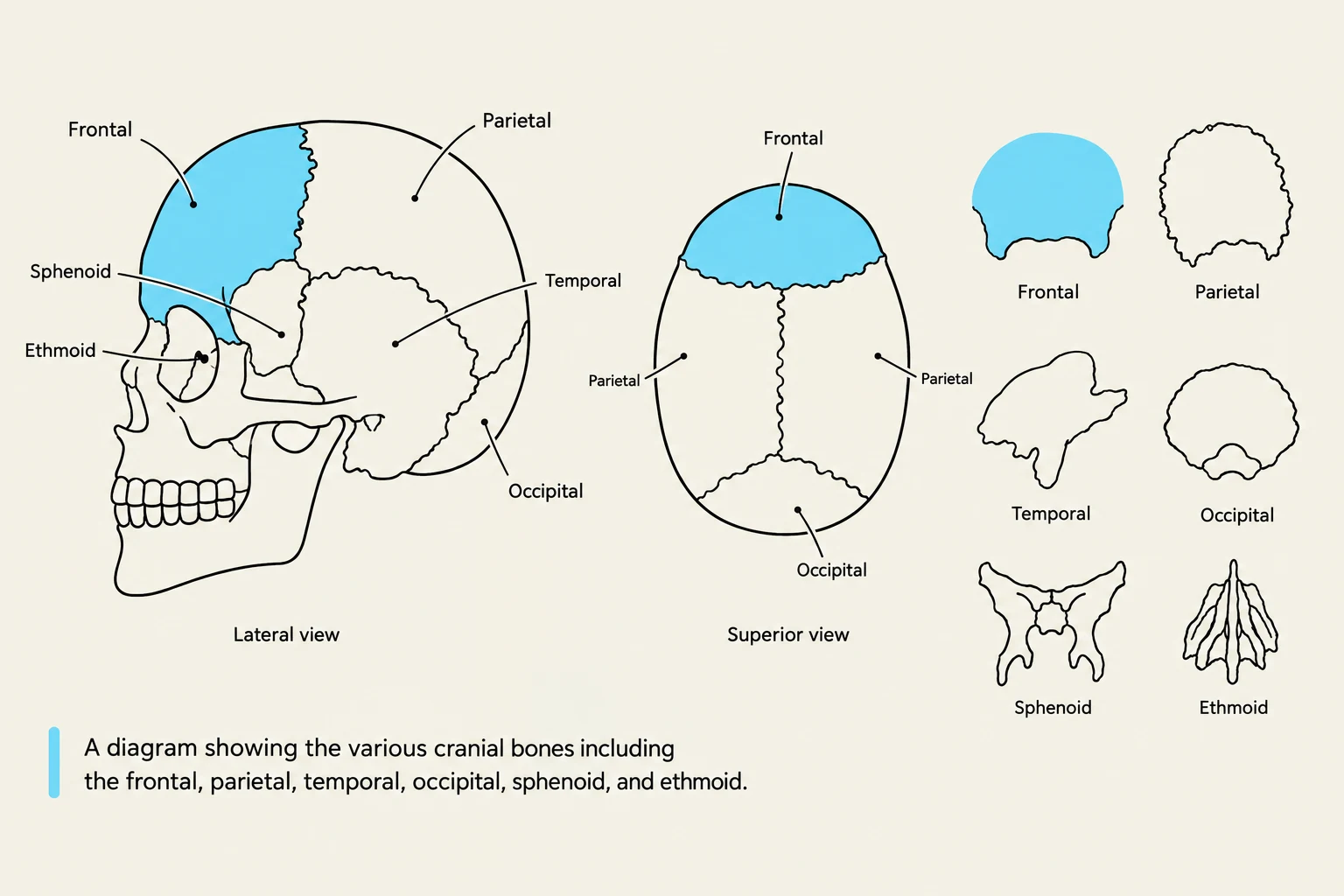
The cranial bones form the "braincase." Each bone has specific landmarks that are critical for anatomical identification:
Frontal bone: Forms the forehead and the superior portion of the orbits. It contains the frontal paranasal sinus.
Parietal bones: Two large bones that form the bulk of the cranial roof. They are joined at the midline by the sagittal suture.
Temporal bones: Located on the lateral sides of the skull. These bones are home to the external acoustic meatus (the ear canal) and the mastoid process.
Occipital bone: Forms the posterior base of the skull. Its most significant feature is the foramen magnum, a large opening that allows the spinal cord to emerge from the brain.
Sphenoid bone: Often described as a complex, butterfly-shaped bone that spans the width of the skull. It contains the sella turcica (which holds the pituitary gland) and the superior orbital fissure.
Ethmoid bone: Situated between the orbits, this bone is essential for the nasal cavity. It features the crista galli (a bony protrusion) and the cribriform plate, which allows olfactory nerves to pass through.
A diagram showing the various cranial bones including the frontal, parietal, temporal, occipital, sphenoid, and ethmoid.
Facial Bones
The facial bones provide the structure for our expressions and the housing for the teeth and nasal passages. Key bones include:
Maxillae: The upper jaw bones that hold the upper teeth.
Mandible: The lower jaw bone. It is unique because it is the only mobile bone of the skull. Key landmarks include the condylar process (which articulates with the temporal bone) and the coronoid process.
Zygomatic bones: Commonly known as the cheekbones.
Nasal bones: Form the bridge of the nose.
Lacrimal bones: Small bones located within the medial orbit.
Palatine bones: Form part of the hard palate (roof of the mouth).
Vomer: A thin bone that forms part of the bony nasal septum.
Exam Tip: Processes vs. Foramina
When studying, remember these definitions: A process is a protrusion or a "point" on a bone (e.g., mastoid process, zygomatic process). A foramen is a hole or opening that allows for the passage of nerves or blood vessels (e.g., foramen magnum, mental foramen).
Cranial Openings and Infant Development
The skull is riddled with foramina (openings) that act as transit points for vital structures. For example, the optic canal allows the optic nerve to reach the eye, while the jugular foramen transmits the jugular vein and artery. The mental foramen is located on the mandible.
In infants, the skull is not a single solid unit. Instead, there are gaps called fontanelles (such as the anterior/bregmatic and posterior/lambdoid fontanelles). These allow the skull to be flexible during birth and provide room for the rapid brain growth that occurs in early childhood.
The Thoracic Cage
The thoracic cage protects the heart and lungs while facilitating the mechanics of breathing. It is composed of the sternum and the ribs, which are connected by costal cartilage.
The Sternum and Ribs
The sternum is the breastbone, consisting of three parts: the manubrium, the body, and the xiphoid process.
The ribs are connected to the sternum via hyaline cartilage (also called costal cartilage), a clear, transparent, and flexible tissue that allows the rib cage to expand during inhalation. Ribs are classified into three distinct types based on their attachment to the sternum:
Vertebrosternal (True) Ribs (1–7): These attach directly to the sternum via their own individual pieces of costal cartilage.
Vertebrochondral (False) Ribs (8–10): These do not attach directly to the sternum; instead, their cartilages fuse with the cartilage of the rib above them.
Vertebral (Floating) Ribs (11–12): These have no anterior attachment to the sternum or costal cartilage at all; they are primarily visible from the posterior side of the body.
The image illustrates the three types of ribs: true ribs (1-7), false ribs (8-10), and floating ribs (11-12), along with a diagram of the thoracic cage showing the manubrium, body, and xiphoid process of the sternum.
An individual rib is characterized by two ends: the sternal end, which faces the sternum, and the vertebral end, which faces the spine to attach to the vertebral facets. The vertebral end features a flat-edged neck to facilitate this connection.
The Vertebral Column
The vertebral column is a series of bones called vertebrae that support the weight of the body and protect the spinal cord. It is divided into five main regions:
Cervical (7 vertebrae): Located in the neck.
Thoracic (12 vertebrae): Located in the chest, articulating with the ribs.
Lumbar (5 vertebrae): Located in the lower back; these have the largest bodies to support significant weight.
Sacral (5 fused vertebrae): Forms the sacrum.
Coccygeal (3-5 fused vertebrae): Forms the coccyx (tailbone).
Specialized Vertebrae: Atlas and Axis
The first two cervical vertebrae are highly specialized to allow for head movement:
C1 (Atlas): Lacks a body and a spinous process. It articulates with the occipital condyles of the skull and allows for the "nodding" motion of the head.
C2 (Axis): Features a unique projection called the dens. The dens fits into the atlas, allowing the head to rotate (the "no" motion).
Regional Differences and Spinal Curvatures
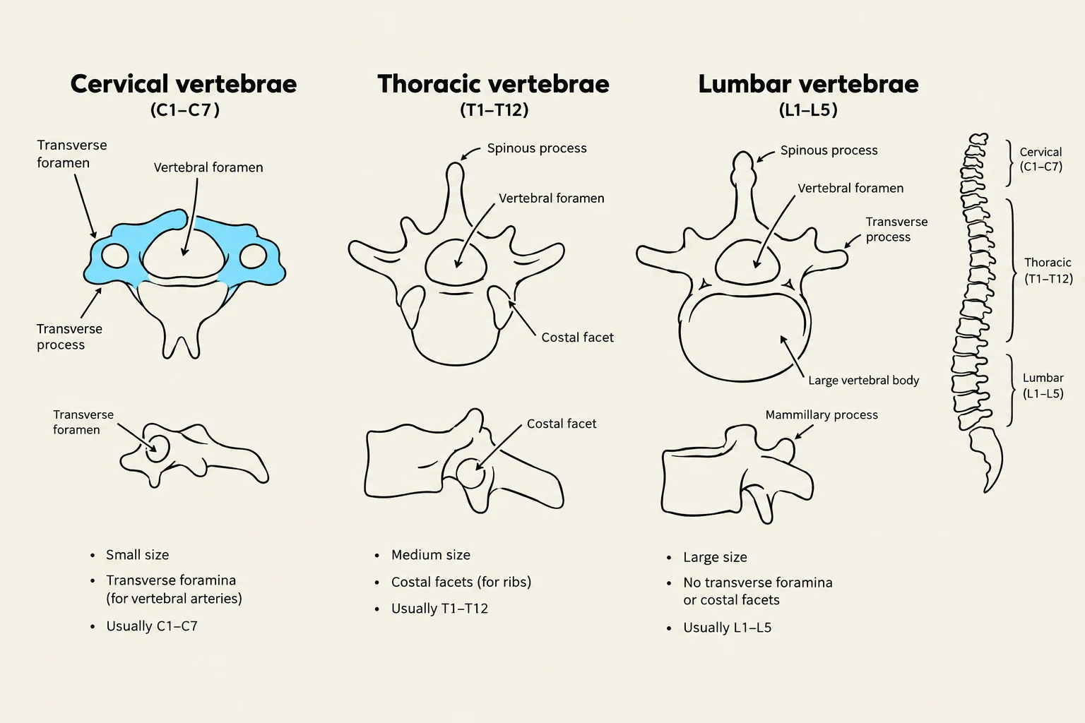
Vertebrae vary significantly by region. For example, cervical vertebrae are small and have transverse foramina (holes in the transverse processes), whereas thoracic vertebrae have long, slender spinous processes and facets for rib attachment. Lumbar vertebrae are massive and blunt to support the trunk.
Diagram illustrating the differences between cervical, thoracic, and lumbar vertebrae, including their anatomical features and typical locations in the spine.
The spine also possesses natural curvatures. Abnormalities in these curves include:
Scoliosis: An abnormal lateral (side-to-side) curvature.
Kyphosis: An exaggerated outward curvature of the thoracic spine (often called a "hunchback").
Lordosis: An exaggerated inward curvature of the lumbar spine.
Clinical Connection: Spinal Injury
In high-impact accidents, such as motor vehicle or motorcycle crashes, the spinal column is at risk. Severe injuries can lead to "internal decapitation," where the connection between the skull and the vertebral column is compromised.
Keep reading for free.
Create an account to finish this reading and make it part of your study routine.