


The human skull undergoes a remarkable transformation from birth through early childhood. Unlike the rigid, fused skull of an adult, an infant's cranium is composed of several distinct bony plates that are not yet fully joined. This anatomical arrangement is a critical evolutionary adaptation, providing the necessary flexibility for the head to pass through the birth canal and allowing the brain to expand rapidly during the first years of life.
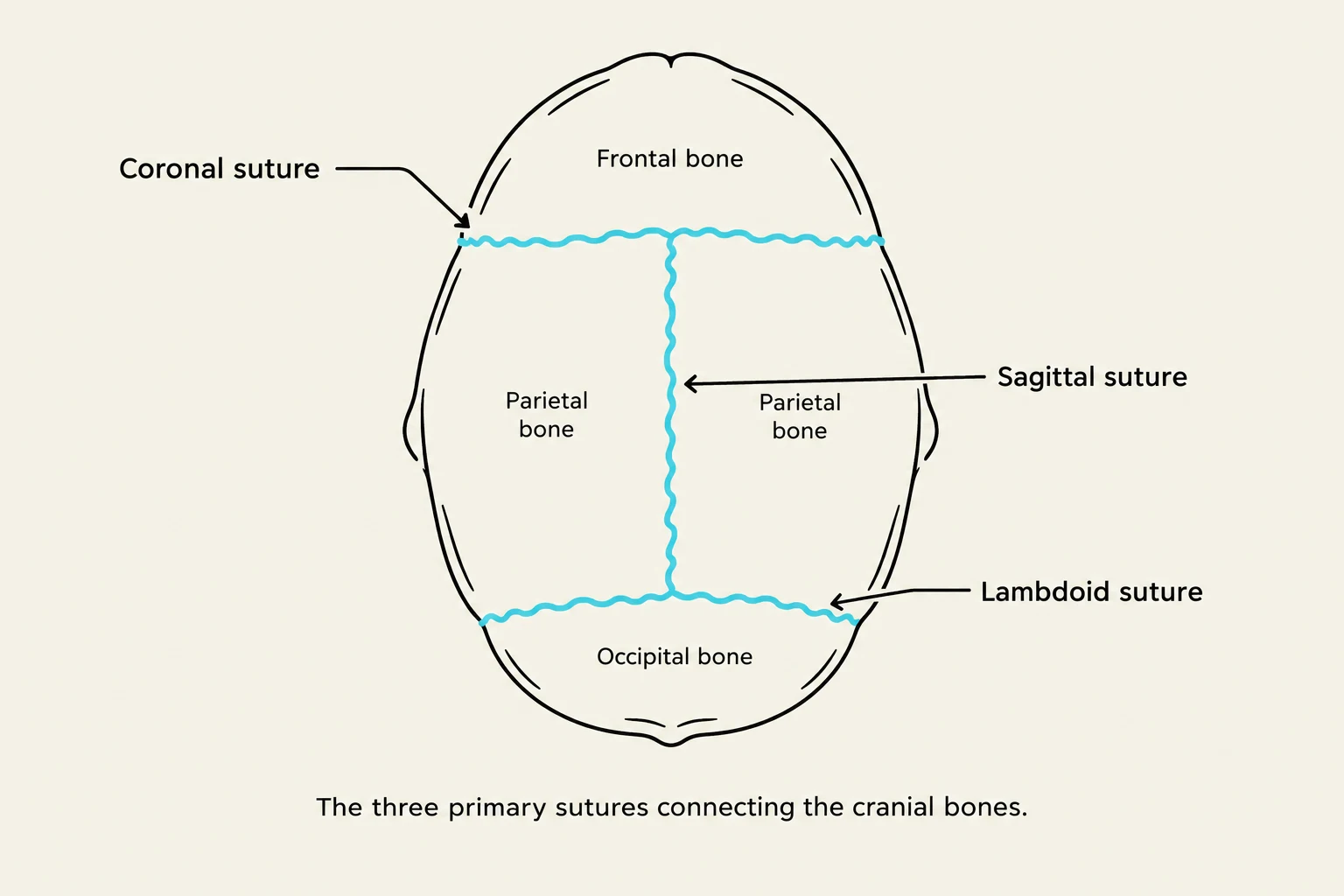
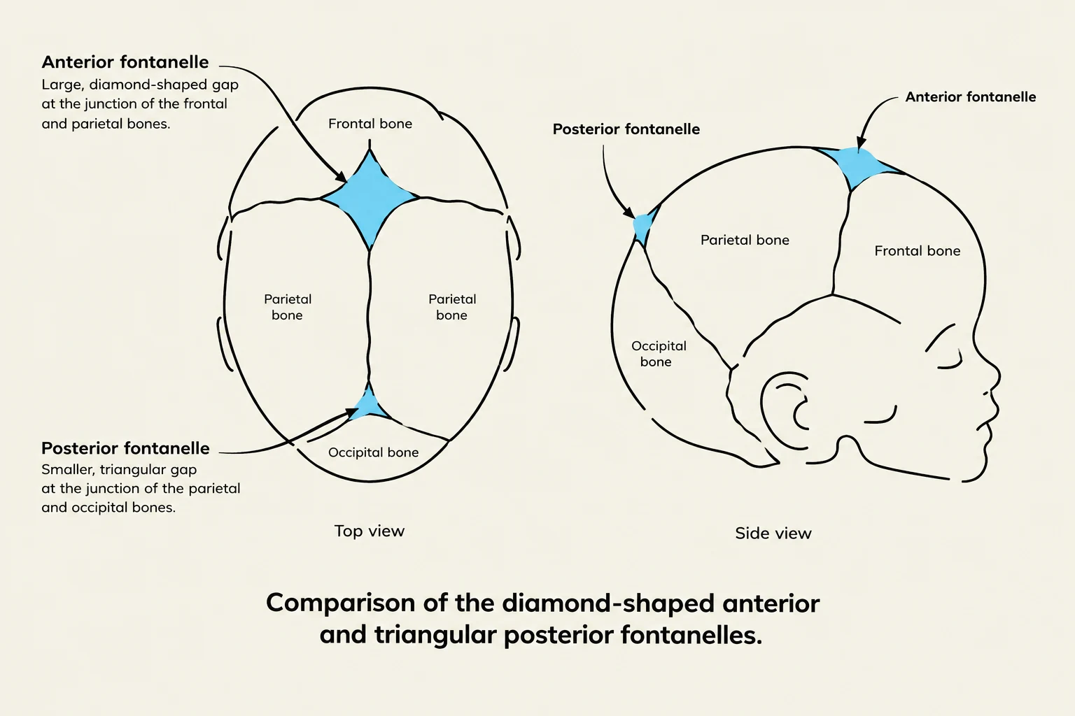
This flexibility is made possible by two specific types of junctions: sutures, which are the fibrous connections between bones, and fontanelles, often referred to as "soft spots," which are non-ossified gaps where multiple sutures meet.
The Cranial Bones: Building Blocks of the Skull
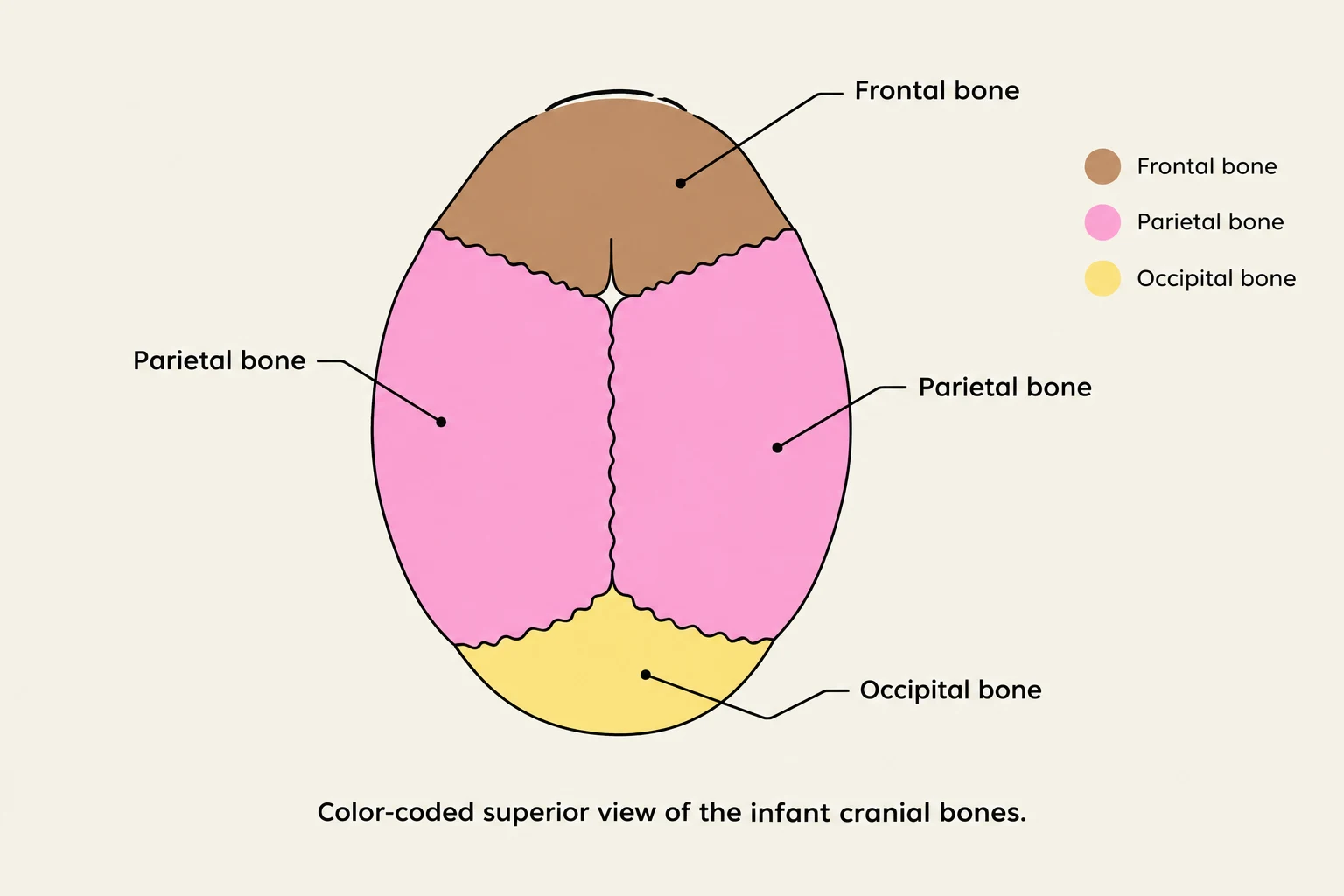
To understand how the skull expands, one must first identify the primary bones that form the cranial vault. When viewed from a superior (top-down) perspective, the skull is organized into specific regions:
- Frontal Bone: Located at the anterior (front/top) portion of the skull.
- Parietal Bones: These are two large, symmetrical bones that occupy the majority of the skull's middle section.
- Occipital Bone: Located at the posterior (back/bottom) portion of the skull.
fontanelles superior view Figure 1: A superior view of the infant skull highlighting the different bone regions and their junctions.
In anatomical diagrams, these bones are often color-coded for clarity. For instance, the frontal bone may be represented in brown, the parietal bones in pink, and the occipital bone in yellow.
Keep reading for free.
Create an account to finish this reading and make it part of your study routine.
Continue reading freeAlready have an account? Log in
Updated