


Mastering the anatomy of the forearm and hand requires more than just memorizing bones; you must understand the complex network of ligaments, nerves, and soft-tissue structures that facilitate fine motor skills and stability. This guide focuses on the "other structures"—the high-yield soft tissues, landmarks, and specialized regions that frequently appear on practical exams and clinical assessments.
Bony Landmarks and Articulations
To orient yourself, always start with the two long bones of the forearm. The radius is located on the lateral side (thumb side), while the ulna is on the medial side (little-finger side).
The Radius and Ulna
The radius includes the head, fovea, neck, radial tuberosity, and the distal styloid process. Proximally, the radial head articulates with the capitulum of the humerus, and distally, it articulates with the scaphoid and lunate bones.
The ulna features the olecranon process (the point of the elbow), the coronoid process, the trochlear notch, the radial notch, the ulnar tuberosity, and the ulnar styloid process. A critical articulation is the trochlear notch, which fits into the trochlea of the humerus. Connecting these two bones is the interosseous membrane, a broad fibrous sheet that spans the space between the radius and ulna.
The Carpal Bones
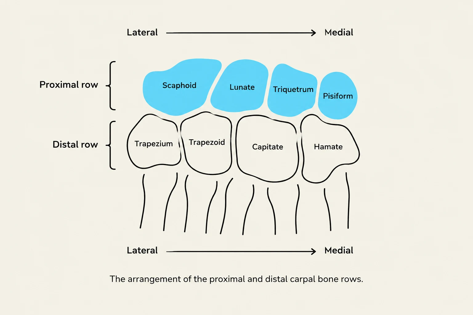
- Proximal Row (Lateral to Medial): Scaphoid, Lunate, Triquetrum, and Pisiform.
- Distal Row (Lateral to Medial): Trapezium, Trapezoid, Capitate, and Hamate.
Study Tip: Mnemonics To remember the proximal row, use: "Scaphoid, Lunate, Triquetrum, Pisiform." For the distal row, use: "Trapezium, Trapezoid, Capitate, Hamate." A common full-circle mnemonic is: "So long to pinky, here comes the thumb" (Scaphoid Lunate Triquetrum Pisiform Hamate Capitate Trapezoid Trapezium).
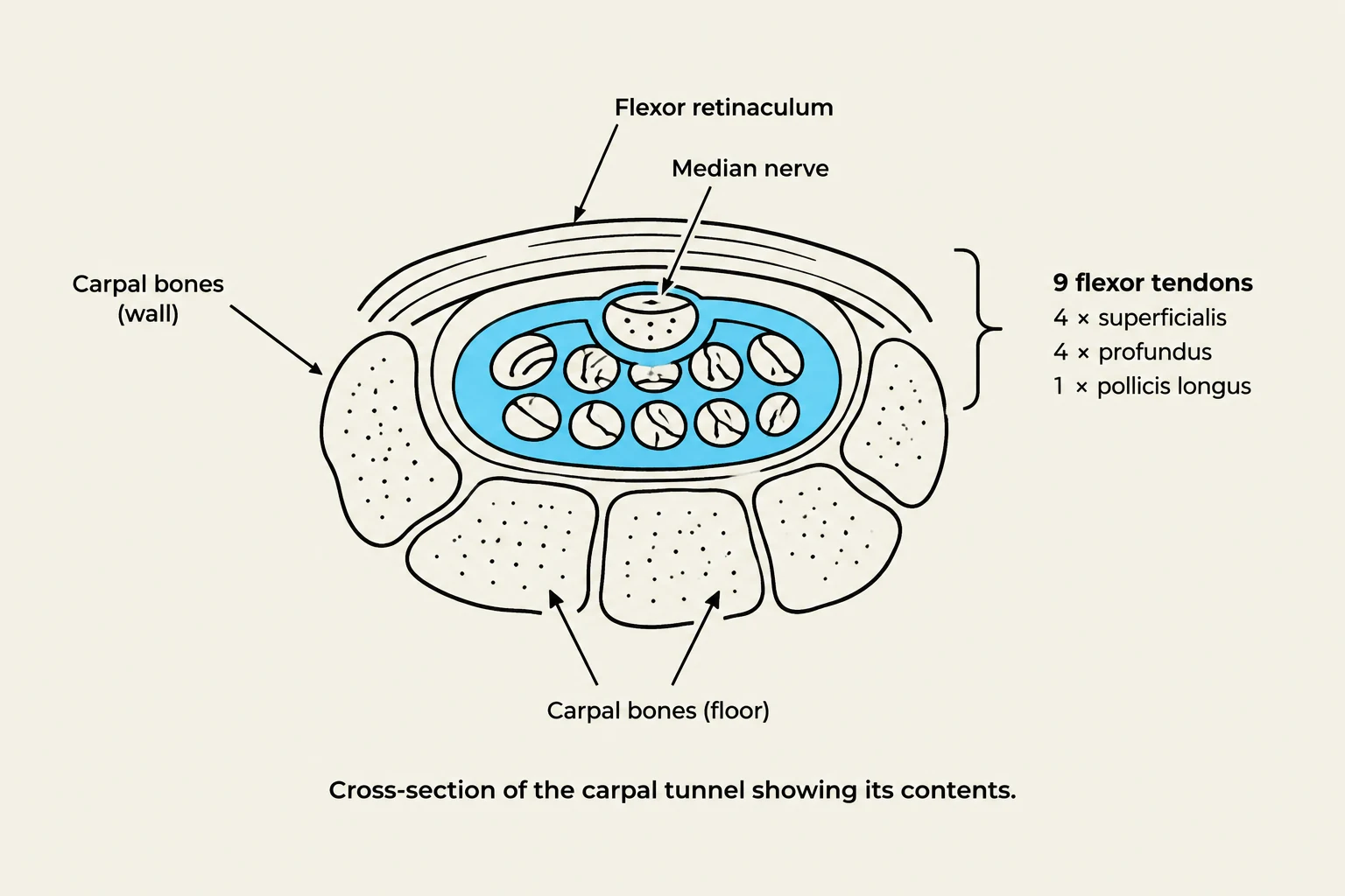
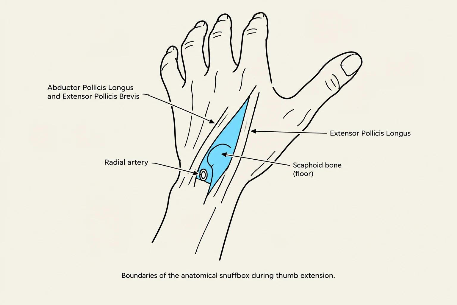
Keep reading for free.
Create an account to finish this reading and make it part of your study routine.
Continue reading freeAlready have an account? Log in
Updated