An overview of the endocrine system, distinguishing between endocrine and exocrine glands. It details the primary endocrine organs (hypophysis, pineal, thyroid, parathyroid, adrenal) and secondary endocrine tissues, along with the anatomy and lymphatic drainage of mammary glands.
The endocrine system is a complex network of glands and organs that work in concert with the nervous system to maintain homeostasis. Unlike the nervous system, which provides rapid-fire electrical signals, the endocrine system utilizes chemical messengers known as hormones. While the effects of hormones are generally slower to manifest, they tend to last much longer than neural impulses.
Endocrine vs. Exocrine Glands
To understand the endocrine system, one must first distinguish it from the exocrine system. The primary difference lies in how they deliver their products.
Endocrine Glands: These glands lack secretory ducts. Instead, they possess an extensive blood supply that allows them to deliver hormones directly into the blood, lymph, or tissue fluid.
Exocrine Glands: These glands release substances through specific openings or ducts onto external body surfaces or into internal body cavities. Common examples include sweat, saliva, tears, digestive juices, and milk.
Exam Tip: If a question asks about the "absence of ducts," it is referring to an endocrine gland. If it mentions "secretory ducts," it is referring to an exocrine gland.
Primary Endocrine Organs and Hormone Synthesis
Hormone synthesis is a fundamental function shared by all endocrine glands. In anatomy, we categorize these into primary endocrine organs and organs that possess endocrine tissue alongside other functions.
Primary Endocrine Organs
The main organs dedicated to endocrine function include:
Hypophysis (Pituitary gland)
Pineal gland (formerly known as the epiphysis)
Thyroid glands
Parathyroid glands
Adrenal glands
Secondary Endocrine Activity
Several organs perform multiple roles, combining endocrine activity with other physiological functions:
Pancreas: Contains endocrine tissue.
Gonads (Ovary/Testis): Contain endocrine tissue.
Placenta: Contains endocrine cells.
Other sites: The kidney, brain, liver, thymus, heart, and gastrointestinal (GI) tract also contain endocrine cells.
The Hypophysis: The Master Gland
The hypophysis, or pituitary gland, is often referred to as the "Master Gland" due to its regulatory role. Anatomically, it is located in the cranial cavity, occupying a central depression in the sella turcica (Turkish saddle) of the basisphenoid bone. This specific depression is known as the hypophyseal fossa.
The hypophysis is composed of two distinct parts with different origins and communication methods with the hypothalamus:
Adenohypophysis (Anterior Pituitary): This is the rostral and ventral portion of the gland. It is connected to the hypothalamus via the hypophyseal portal vascular system. It produces several vital hormones, including:
Growth hormone
Thyroid-stimulating hormone (TSH)
Adrenocorticotropic hormone (ACTH)
Follicle-stimulating hormone (FSH)
Luteinizing hormone (LH)
Prolactin
Neurohypophysis (Posterior Pituitary): This part is an extension of the encephalon (specifically the hypothalamus). It is connected to the hypothalamus by a neural stem. It stores and releases:
Oxytocin
Vasopressin (also known as antidiuretic hormone)
The Pineal Gland
Located in the middle of the encephalon within the area known as the diencephalon, the pineal gland is found in a caudo-dorsal position relative to the thalamus. Its shape is reminiscent of a pine cone.
The primary function of the pineal gland is the production of melatonin. Melatonin is a critical hormone that modulates sleep patterns by regulating both circadian (daily) and seasonal cycles.
Thyroid and Parathyroid Glands
Thyroid Glands
The thyroid glands are responsible for producing thyroxine hormones, specifically T3 and T4. In most dogs, the thyroid is a paired gland consisting of a right and left lobe. These lobes lie lateral to the trachea, caudal to the larynx, and may sometimes overlap the larynx. Occasionally, the two lobes are connected by a structure called the isthmus.
Parathyroid Glands
The parathyroid glands produce parathyroid hormones. There are normally four of these glands (two on each side). In species like dogs and cats, they are typically embedded within the thyroid gland tissue.
Clinical Note: During dissection, parathyroid glands can be distinguished from thyroid tissue by their color. The parathyroid glands appear pale, whereas the thyroid gland has a distinct "red-brick" color.
Adrenal Glands
The adrenal glands are described as "small, but MIGHTY" due to their significant physiological impact. They are located in a retroperitoneal position, situated craniomedially to the cranial pole of the kidneys, and are often hidden by fat.
The adrenal gland is organized into two main functional layers: the cortex and the medulla. They produce several critical hormones:
Cortisol: Affects muscle, bone, skin, the immune system, the vascular system, the CNS, the liver, and the kidneys.
Aldosterone (Mineralocorticoid): Regulates mineral balance.
Epinephrine (Adrenaline): Involved in the stress response.
Androgens.
Mammary Glands and Lymphatic Drainage
While the mammary glands are technically exocrine glands (secreting milk through ducts), they are a vital part of reproductive anatomy. They are subcutaneous, enlarged sweat glands that produce colostrum and milk.
Anatomy and Distribution
Mammary glands develop along mammary ridges (milk lines) that extend from the axilla (armpit) to the inguinal (groin) regions. Each gland is separated by connective tissue septa and secretes via a teat or papilla.
The number of glands varies by species:
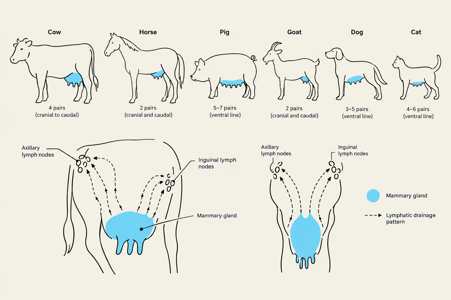
Dogs: Normally five pairs, with 10–12 openings per gland.
Cats: Normally four pairs, with 4–8 openings per gland
This diagram illustrates the mammary gland and lymphatic drainage patterns in various domestic animals, including cows, horses, pigs, goats, dogs, and cats. It shows the location of axillary and inguinal lymph nodes relative to the mammary glands.
Lymphatic Drainage
Understanding the lymphatic drainage of the mammary glands is clinically essential, particularly regarding the metastasis of mammary tumors. The drainage follows specific pathways:
Cranial/Thoracic region: Drains via the axillary and accessory axillary lymph nodes.
Abdominal/Inguinal region: Drains via the superficial inguinal lymph nodes.
The lymphatic vessels eventually lead toward major trunks such as the lumbar lymphatic trunk, the visceral lymphatic trunk, and the cisterna chyli, ultimately reaching the thoracic duct.
Keep reading for free.
Create an account to finish this reading and make it part of your study routine.