Anatomy of Dicotyledonous and Monocotyledonous Plants
Explore the fundamental tissue systems of plants and the distinct anatomical differences in roots, stems, and leaves between dicotyledonous and monocotyledonous species, focusing on vascular bundle organization and secondary growth.
The study of the internal structure of plants is known as anatomy. While all flowering plants share certain fundamental tissue systems, there are profound structural differences between dicotyledonous (dicot) and monocotyledonous (monocot) species. These differences are most visible when examining the organization of the vascular system within the roots, stems, and leaves.
The Fundamental Tissue Systems
To understand plant anatomy, we must first categorize tissues based on their location and function. Plant tissues are broadly classified into meristematic tissues (responsible for growth) and permanent tissues (specialized for specific functions). These permanent tissues are organized into three distinct systems:
Epidermal Tissue System: The outermost protective covering, comprising epidermal cells, stomata, and epidermal appendages like hairs or trichomes.
Ground Tissue System: Everything except the epidermis and vascular bundles. It includes the cortex, pericycle, and pith, and is composed of simple tissues like parenchyma, collenchyma, and sclerenchyma.
Vascular Tissue System: The conducting system responsible for transporting water, minerals, and food.
The Vascular Tissue System and Bundles
The vascular system is composed of complex tissues: xylem (which conducts water and minerals) and phloem (which conducts photosynthates). Together, these tissues form vascular bundles.
The arrangement of these bundles varies significantly:
Radial Arrangement: Xylem and phloem are arranged alternately along different radii. This is a characteristic feature of roots.
Conjoint Arrangement: Xylem and phloem are situated on the same radius. This is common in stems and leaves.
A critical distinction in conjoint bundles is the presence of cambium.
Open Vascular Bundles: Found in dicot stems, these contain cambium between the xylem and phloem, allowing for secondary growth (increase in girth).
Closed Vascular Bundles: Found in monocots, these lack cambium and therefore cannot undergo secondary growth.
Diagrams illustrating plant vascular tissues: (a) cross-section of a vascular bundle, (b) longitudinal view of xylem and phloem, and (c) longitudinal view showing xylem, phloem, and cambium.
Figure 6.2 Various types of vascular bundles : (a) radial (b) conjoint closed (c) conjoint open
Root Anatomy: Dicot vs. Monocot
The root serves as the primary anchor and water-absorber. Both types share a basic structure including the epiblema (epidermis), cortex, endodermis, pericycle, and stele (the central cylinder containing the pith and vascular tissues).
Dicotyledonous Roots
In a dicot root, the epiblema often features unicellular root hairs for absorption. The cortex consists of several layers of parenchyma. The innermost layer of the cortex is the endodermis, which contains Casparian strips—waxy, water-impermeable deposits of suberin. The pericycle, located just inside the endodermis, is responsible for the initiation of lateral roots. Dicot roots typically have a small or inconspicuous pith and fewer xylem bundles.
Monocotyledonous Roots
While the general layers are similar, monocot roots are distinguished by a polyarch condition, meaning they possess more than six xylem bundles. Additionally, the pith in monocot roots is large and well-developed. Unlike dicots, monocot roots do not undergo secondary growth.
Diagrams illustrating the cross-section of a plant root, showing different tissue layers such as epidermis, cortex, endodermis, pericycle, xylem, and phloem.
Figure 6.3 T.S. : (a) Dicot root (Primary) (b) Monocot root
Stem Anatomy: Ring vs. Scattered
The stem provides structural support and transport pathways. The arrangement of vascular bundles here is one of the easiest ways to distinguish the two groups.
Dicotyledonous Stems
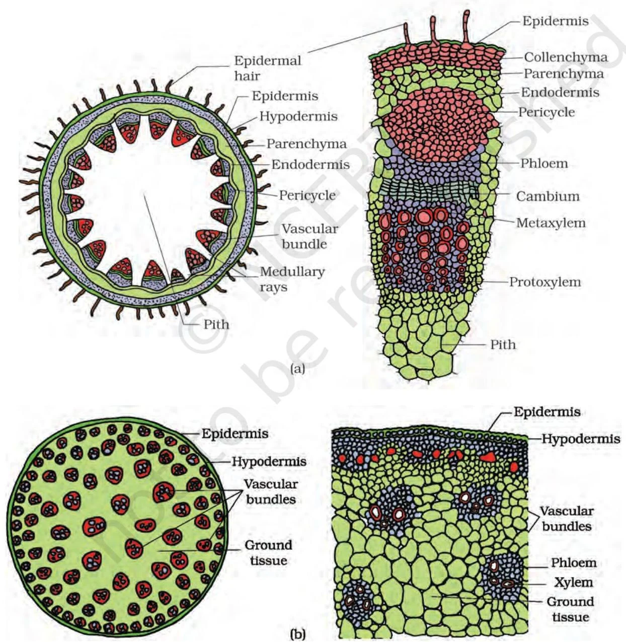
Dicot stems are characterized by a ring arrangement of vascular bundles. The cortex includes an outer hypodermis made of collenchyma cells to provide mechanical strength. The endodermis in dicot stems often acts as a starch sheath due to its richness in starch grains. Between the vascular bundles, radially placed parenchymatous cells form medullary rays.
Monocotyledonous Stems
In contrast, monocot stems feature scattered vascular bundles throughout the ground tissue. Each bundle is typically surrounded by a sclerenchymatous bundle sheath. A key identifying feature is the absence of phloem parenchyma and the presence of water-containing cavities within the vascular bundles.
Diagrams illustrating the cross-section and longitudinal section of plant stems, showing various tissues like epidermis, vascular bundles, and pith.
Figure 6.4 T.S. of stem : (a) Dicot (b) Monocot
Leaf Anatomy: Dorsiventral vs. Isobilateral
Leaves are specialized organs for photosynthesis, and their internal tissue, the mesophyll, reflects their venation patterns.
Dorsiventral (Dicot) Leaves
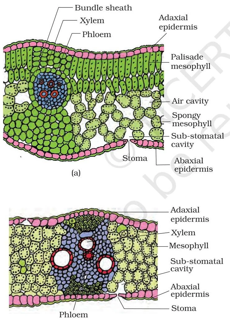
Dicot leaves show a clear distinction between the upper (adaxial) and lower (abaxial) surfaces. The mesophyll is differentiated into two distinct regions:
Palisade parenchyma: Elongated cells arranged vertically near the upper epidermis.
Spongy parenchyma: Loosely arranged, oval cells with large air spaces located below the palisade layer.
These leaves typically exhibit reticulate venation (net-like).
Isobilateral (Monocot) Leaves
Monocot leaves are often isobilateral, meaning they look similar on both sides. Stomata are usually present on both surfaces, and the mesophyll is not differentiated into palisade and spongy layers. They exhibit parallel venation.
Biological Adaptation: Bulliform Cells
In many grasses (monocots), specialized large, empty epidermal cells called bulliform cells are present on the upper surface. When water is abundant, these cells are turgid and the leaf remains flat. During water stress, they become flaccid, causing the leaf to curl inwards to minimize water loss through transpiration.
Diagrams illustrating the cross-section of a plant leaf, showing different tissue layers and structures such as epidermis, mesophyll, xylem, phloem, and stomata.
Figure 6.5 T.S. of leaf : (a) Dicot (b) Monocot
Summary Table for Quick Revision
Feature
Dicotyledonous
Monocotyledonous
Root Vascular Bundles
Fewer; small pith
Polyarch (>6); large pith
Stem Vascular Bundles
Arranged in a ring; Open
Scattered; Closed
Stem Hypodermis
Collenchymatous
Sclerenchymatous
Leaf Mesophyll
Differentiated (Palisade & Spongy)
Undifferentiated
Leaf Venation
Reticulate
Parallel
Exam Tip: If a question asks you to identify a plant stem based on a transverse section, look first at the vascular bundles. If they are in a neat ring, it is a dicot; if they are scattered like stars in a sky, it is a monocot.
Keep reading for free.
Create an account to finish this reading and make it part of your study routine.