This text describes membranous and non-membraneous organelles including the endoplasmic reticulum, ribosomes, Golgi apparatus, lysosomes, proteasomes, peroxisomes, mitochondria, and the nucleus.
The cell is a complex, highly organized unit of life, functioning much like a miniature factory. Within the cell, various specialized structures called organelles carry out specific tasks necessary for survival, growth, and reproduction. These organelles are suspended in a gel-like substance known as the cytoplasm, which includes a watery intracellular fluid called cytosol. To understand how a cell functions, we must distinguish between two primary categories of organelles: those enclosed by membranes and those that are not.
Membranous Organelles
Membranous organelles are essentially sacs or canals constructed from the same phospholipid bilayer material that makes up the plasma membrane. These membranes allow the cell to create distinct internal environments for different chemical reactions.
The Nucleus: The Control Center
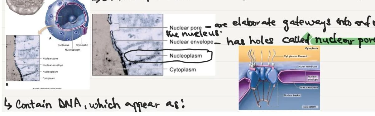
The nucleus is a large, spherical membranous structure typically located near the center of the cell. It serves as the repository of genetic information, containing DNA which determines both the structure and function of the cell, as well as heredity.
The nucleus is enclosed by the nuclear envelope, which surrounds the internal fluid known as nucleoplasm. This envelope is not a solid wall; rather, it is perforated by numerous holes called nuclear pores that regulate the passage of materials. Inside the nucleus, DNA can be seen as chromatin threads or granules in non-dividing cells, or as distinct chromosomes during the early stages of cell division.
Diagrams illustrating the structure of the cell nucleus, including nuclear pores and their function as gateways.
Endoplasmic Reticulum (ER) and Protein Synthesis
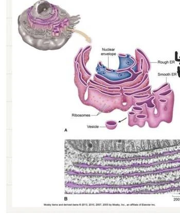
The endoplasmic reticulum is a vast network of membranous-walled canals and flat, curving sacs arranged in parallel rows throughout the cytoplasm. It extends from the plasma membrane all the way to the nucleus and plays a critical role in intracellular transport.
The ER is divided into two distinct types:
Rough ER: This section is "studded" with ribosomes on its outer surface. These ribosomes are the sites of protein synthesis. Proteins made on the rough ER are typically destined for export or for use in membranes.
Smooth ER: This section lacks ribosomes on its surface. Its functions are more varied, including the synthesis of certain lipids and carbohydrates, the creation of membranes, and the removal and storage of calcium ions (Ca2+) from the cell's interior.
Diagrams illustrating the structure of the endoplasmic reticulum and its relationship with the nuclear envelope, including rough ER, smooth ER, ribosomes, and vesicles.
The Golgi Apparatus and Lysosomes
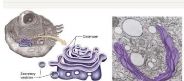
Once proteins are synthesized in the ER, they are sent to the Golgi apparatus for processing. The Golgi consists of flattened sacs called cisternae stacked upon one another near the nucleus. It acts as a processing and packaging center; proteins leave the final cisterna in a vesicle, which may then secrete the contents outside the cell.
Diagram of the Golgi apparatus, showing cisternae and secretory vesicles, alongside a micrograph of a similar cellular structure.
From the Golgi apparatus, small membranous sacs called lysosomes can pinch off. Lysosomes function as the cell's digestive system, containing enzymes that break down defective cell parts, such as damaged integral membrane proteins or trapped particles.
Mitochondria and Peroxisomes
Mitochondria are often referred to as the "powerhouses" of the cell. They consist of a double membrane (an inner and outer membrane) separated by fluid. Thousands of enzyme molecules attached to these membranes catalyze oxidation reactions that provide almost all of the cell's energy supply. Interestingly, mitochondria contain their own DNA, allowing them to produce their own enzymes and replicate themselves.
Diagrams illustrating the structure and function of mitochondria and the nucleus within a cell.
Peroxisomes are smaller membranous sacs that contain enzymes specifically designed to detoxify harmful substances that enter the cell. They are particularly abundant in liver and kidney cells.
Non-membraneous Organelles
Non-membraneous organelles are not enclosed by a lipid bilayer; instead, they are composed of microscopic filaments or other specialized protein structures.
Ribosomes
Ribosomes are the primary machines of protein synthesis. They are made of two pieces—a large subunit and a small subunit—each composed of RNA and protein. Ribosomes can be found in two states:
Attached to the Rough ER: These produce proteins intended for export.
Free in the Cytoplasm: These produce proteins for the cell's own "domestic" use.
Proteasomes
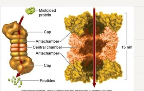
While lysosomes digest large structures, proteasomes are protein cylinders found throughout the cytoplasm that handle smaller tasks. They are responsible for breaking down abnormal or misfolded proteins, as well as normal proteins that the cell no longer requires.
Diagram illustrating the mechanism of a proteasome, a cellular machine that breaks down proteins.
Study Tip: Think of the difference between Lysosomes and Proteasomes as "Bulk Waste Management" vs. "Precision Recycling." Lysosomes handle large, complex debris, while proteasomes target specific, individual protein molecules.
The Cytoskeleton and Cellular Support
The cytoskeleton provides the internal framework of the cell, supporting its shape and allowing for movement. It is composed of a three-dimensional lattice of fibers of varying lengths.
The cytoskeleton includes:
Microfilaments: The smallest fibers, which can act as "cellular muscles" by sliding past each other to cause cell shortening.
Intermediate Filaments: Twisted protein strands that provide structural support.
Microtubules: The thickest fibers, made of protein subunits arranged in a spiral, which function to move things around inside the cell.
The centrosome is a non-membranous structure (also called the microtubule organizing center) that coordinates the building and breaking apart of these microtubules.
Summary Table of Organelle Functions
Organelle
Type
Primary Function
Nucleus
Membranous
Stores DNA; controls cell activities
Rough ER
Membranous
Protein synthesis (via attached ribosomes)
Smooth ER
Membranous
Lipid synthesis; Ca2+ storage
Golgi Apparatus
Membranous
Processing and packaging proteins
Lysosomes
Membranous
Digestion of defective cell parts
Mitochondria
Membranous
Energy (ATP) production
Ribosomes
Non-membranous
Protein synthesis
Proteasomes
Non-membranous
Degradation of misfolded proteins
Peroxisomes
Membranous
Detoxification of harmful substances
Keep reading for free.
Create an account to finish this reading and make it part of your study routine.