


The knee joint is one of the most complex and highly loaded structures in the human body. It serves as a critical link in the kinetic chain, facilitating movement while simultaneously managing significant compressive and shear forces. To understand how the knee moves, how it fails under stress, and how it is rehabilitated, one must first master its anatomy. This article explores the intricate relationship between the bones, bony landmarks, ligaments, and other inert soft tissues that define the knee's structural integrity.
The Skeletal Framework
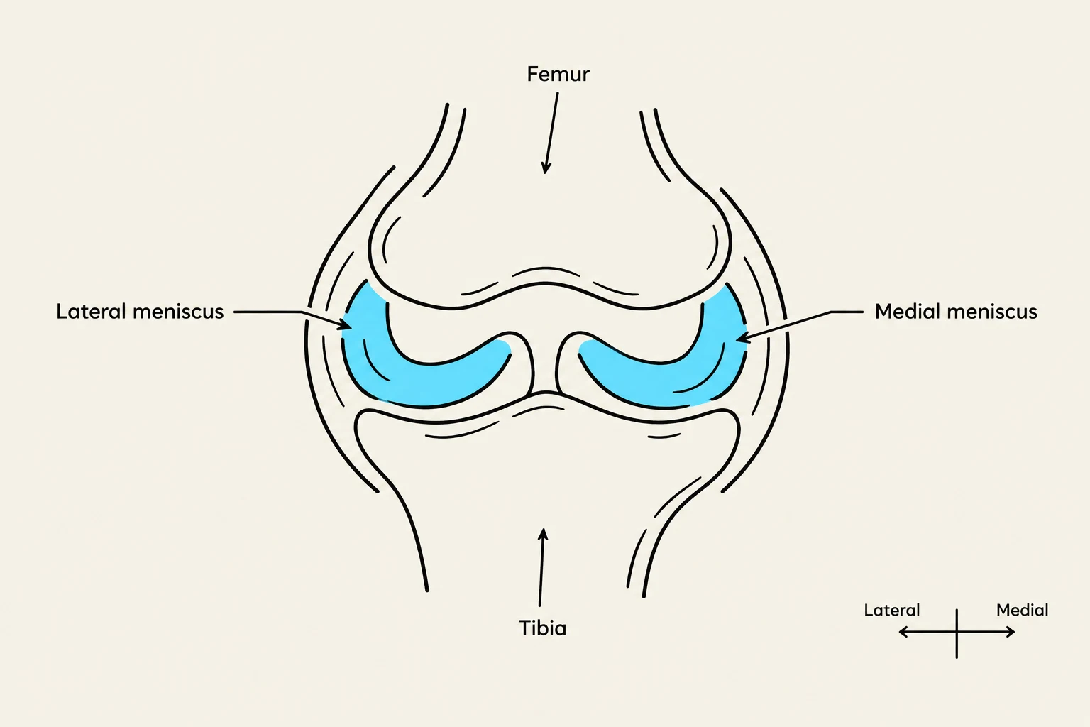
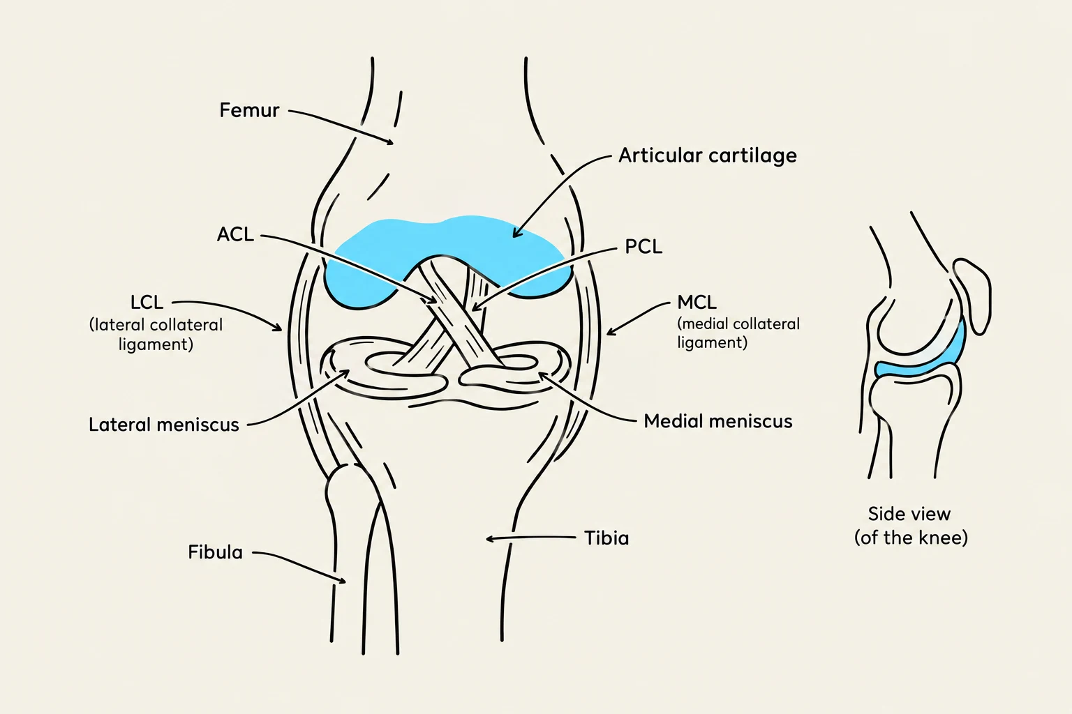
The foundation of the knee joint is built upon four primary bones: the femur, the tibia, the fibula, and the patella.
- Femur: The distal end of the thigh bone forms the upper portion of the joint.
- Tibia: The larger of the two lower leg bones, which bears the majority of the body's weight.
- Fibula: Located laterally to the tibia, the fibula does not directly participate in the weight-bearing aspect of the knee joint but serves as a vital attachment point for various ligaments and muscles.
- Patella: Known as the kneecap, this sesamoid bone sits within the quadriceps tendon, increasing the mechanical advantage of the extensor muscles.
Bony Landmarks and Surface Topography
Bony landmarks are essential for clinical assessment, as they serve as attachment points for muscles and ligaments and define the joint's geometry.
On the femur, we find the lateral epicondyle, which serves as a key attachment site for collateral ligaments. Moving down the shaft of the femur, the linea aspera acts as a prominent ridge for muscle attachment. Near the distal end of the femur, the medial supracondylar ridge and the lateral supracondylar ridge provide structural boundaries and attachment points for soft tissues.
Keep reading for free.
Create an account to finish this reading and make it part of your study routine.
Continue reading freeAlready have an account? Log in
Updated