The human brain is one of the most complex organs in the body, serving as the center for conscious thought, sensory perception, and the regulation of vital autonomic functions. To function effectively, the brain requires a highly specialized environment that provides physical stability, chemical regulation, and protection from mechanical injury. This article explores the anatomical organization of the cerebrum and the three primary defense mechanisms that support it: the cranial meninges, cerebrospinal fluid (CSF), and the blood-brain barrier (BBB).
Organization of Neural Tissue
The brain's functional capacity is determined by the organization of its neural tissues, which are categorized into two distinct types: grey matter and white matter.
Grey Matter and White Matter
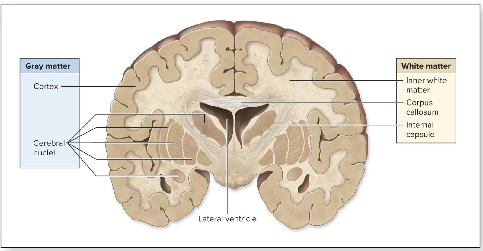
Grey matter consists of neuron and interneuron cell bodies, dendrites, unmyelinated axons, and terminal arborizations. In the cerebrum, the outer layer of grey matter is known as the cerebral cortex. Within the deeper white matter of the cerebrum, grey matter forms discrete clusters known as cerebral nuclei (or basal nuclei), which are involved in various functions such as movement and emotion.
White matter is located deep to the cerebral cortex and derives its color from the high concentration of myelin surrounding the axons. These myelinated axons are organized into bundles called tracts. In the cerebrum, these include:
Association tracts: Connect areas within a single hemisphere.
Commissural tracts: Connect the left and right hemispheres (e.g., the corpus callosum).
Projection tracts: Connect the cerebrum to lower brain regions or the spinal cord (e.g., the internal capsule).
This diagram illustrates a coronal view of the human brain, highlighting the distinctions between gray matter (cortex and cerebral nuclei) and white matter (inner white matter, corpus callosum, and internal capsule), along with the lateral ventricle.
The Cerebrum: Structure and Lobes
The cerebrum is the seat of intellectual functions, including memory, judgment, and voluntary motor control. Its surface is not smooth; instead, it is characterized by folds called gyri (ridges) and shallow depressions called sulci (grooves). Deeper grooves are referred to as fissures.
The cerebrum is divided into two cerebral hemispheres by the longitudinal fissure. Each hemisphere is further subdivided into five functional lobes:
Frontal lobe: Responsible for voluntary movement, decision making, and personality.
Parietal lobe: Involved in general sensory functions like touch and spatial awareness.
Temporal lobe: Primarily involved with hearing and smell.
Insula: Located deep to the lateral sulcus, involved in taste and emotion.
Cranial Meninges
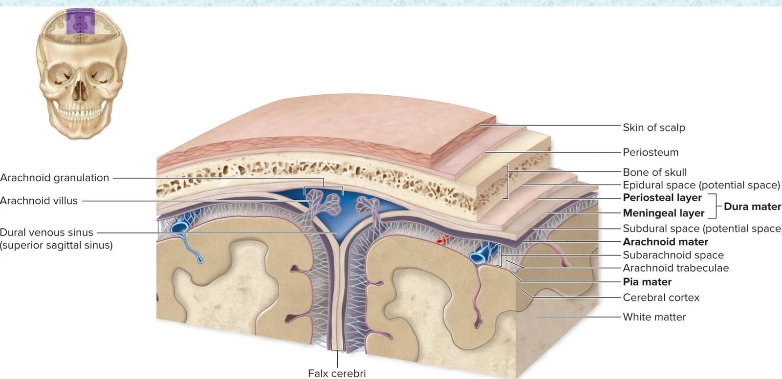
To protect the soft neural tissue from the hard bones of the skull, the brain is enveloped by three layers of connective tissue known as the cranial meninges.
The Three Layers
From the innermost layer to the outermost, the meninges consist of:
Pia mater: A thin, delicate, and highly vascularized layer that adheres tightly to every contour of the brain's surface.
Arachnoid mater: The middle layer, named for its spider-web-like appearance caused by the arachnoid trabeculae (collagen and elastic fibers) that extend into the subarachnoid space.
Dura mater: The toughest, outermost layer composed of dense irregular connective tissue. In the cranium, it consists of a superficial periosteal layer (attached to the skull) and a deeper meningeal layer.
This diagram illustrates the layers of the meninges surrounding the brain, including the dura mater, arachnoid mater, and pia mater, as well as associated structures like the dural venous sinus and falx cerebri.
Dural Septa and Venous Sinuses
The meningeal layer of the dura mater extends into the cranial cavity to form cranial dural septa, which partition and support the brain. Key septa include the falx cerebri (separating the cerebral hemispheres) and the tentorium cerebelli (separating the cerebrum from the cerebellum).
Where the two layers of the dura mater separate, they form large, blood-filled spaces called dural venous sinuses. These sinuses, such as the superior sagittal sinus and transverse sinuses, drain blood from the brain toward the internal jugular veins.
Cerebrospinal Fluid (CSF) and the Ventricular System
Cerebrospinal fluid (CSF) is a clear, colorless liquid that circulates within the brain's cavities and the subarachnoid space.
Functions of CSF
CSF provides three vital life-support functions:
Buoyancy: It supports more than $95%$ of the brain's weight, preventing the organ from being crushed by its own mass.
Protection: It acts as a liquid cushion/buffer against sudden physical movements.
Environmental stability: It transports nutrients and removes metabolic waste while protecting neurons from chemical fluctuations.
Production and Circulation
CSF is produced by the choroid plexus, a structure composed of ependymal cells and capillaries located within the brain's ventricles. The circulation follows a specific pathway:
CSF is produced in the lateral ventricles.
It flows through the interventricular foramen into the third ventricle.
It moves through the cerebral aqueduct into the fourth ventricle.
From the fourth ventricle, it enters the subarachnoid space via the lateral and median apertures.
Excess CSF is eventually expelled from the subarachnoid space through arachnoid villi (and larger arachnoid granulations) into the dural venous sinuses.
Clinical Note: Hydrocephalus
If CSF production exceeds the rate of removal or drainage, the fluid can accumulate, leading to a condition called hydrocephalus. This causes increased intracranial pressure and brain distortion, often requiring the surgical implantation of a shunt to drain the excess fluid.
This illustration shows the flow of cerebrospinal fluid (CSF) through the brain's ventricles and subarachnoid space, highlighting its production by the choroid plexus and reabsorption into the dural venous sinuses via arachnoid villi.
The Blood-Brain Barrier (BBB)
The blood-brain barrier (BBB) is a highly selective physiological barrier that regulates the entry of substances from the blood into the brain's interstitial fluid.
The barrier is maintained by a combination of:
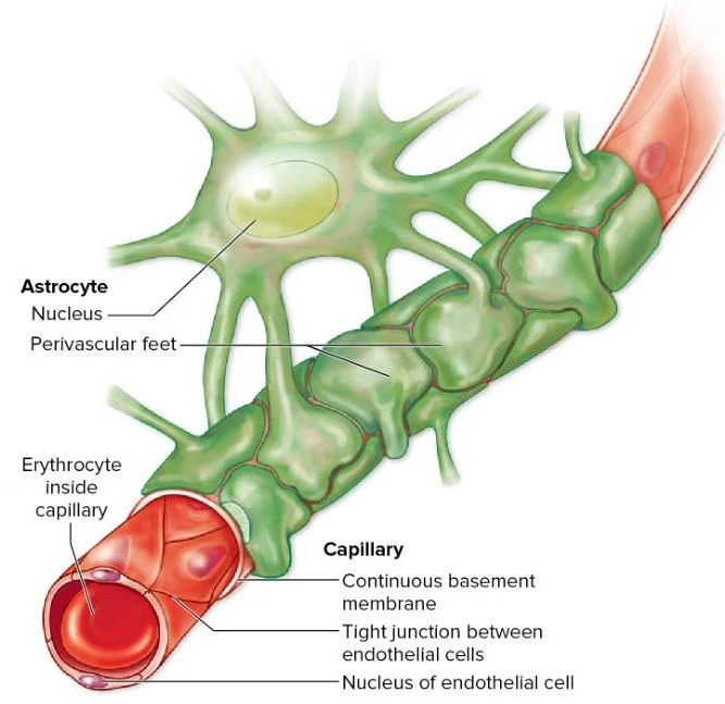
Capillary endothelial cells joined by tight junctions.
A continuous basement membrane.
Astrocyte perivascular feet that wrap around the capillaries to act as "gatekeepers".
Because of this barrier, only lipid-soluble compounds (such as alcohol or nicotine) can easily diffuse across. However, the BBB is intentionally reduced or absent in specific areas to allow for essential functions, such as the choroid plexus (to allow CSF production) and the hypothalamus and pineal gland (to allow hormones to access the blood).
Illustration of an astrocyte interacting with a capillary, showing an erythrocyte within the capillary and details of the capillary wall.
Keep reading for free.
Create an account to finish this reading and make it part of your study routine.