Anatomy and Physiology of the Spinal Cord and Spinal Nerves
Explore the structure and function of the spinal cord, including its gross anatomy, meninges, internal organization of gray and white matter, spinal nerves, plexuses, and reflex arcs. This content also covers the sympathetic division of the autonomic nervous system.
The spinal cord serves as the vital communication bridge between the brain and the rest of the body. It functions as a conduit for sensory information traveling upward to the brain and motor commands traveling downward to muscles and glands. Beyond simple transmission, the spinal cord is also the center for rapid, involuntary responses known as reflexes.
Gross Anatomy and Spinal Cord Regions
The spinal cord is approximately 16–18 inches long and about ¾ of an inch in diameter. It begins at the foramen magnum and extends inferiorly to the level of the L1 vertebra. Because the spinal cord is shorter than the vertebral canal that houses it, it does not extend the full length of the spine.
The spinal cord is divided into five distinct regions, named according to the spinal nerves that attach to them:
Cervical part
Thoracic part
Lumbar part
Sacral part
Coccygeal part
As the cord travels downward, it undergoes changes in diameter. You will find a cervical enlargement in the inferior cervical part that innervates the upper limbs, and a lumbosacral enlargement that innervates the lower limbs.
The Inferior End: Conus Medullaris and Cauda Equina
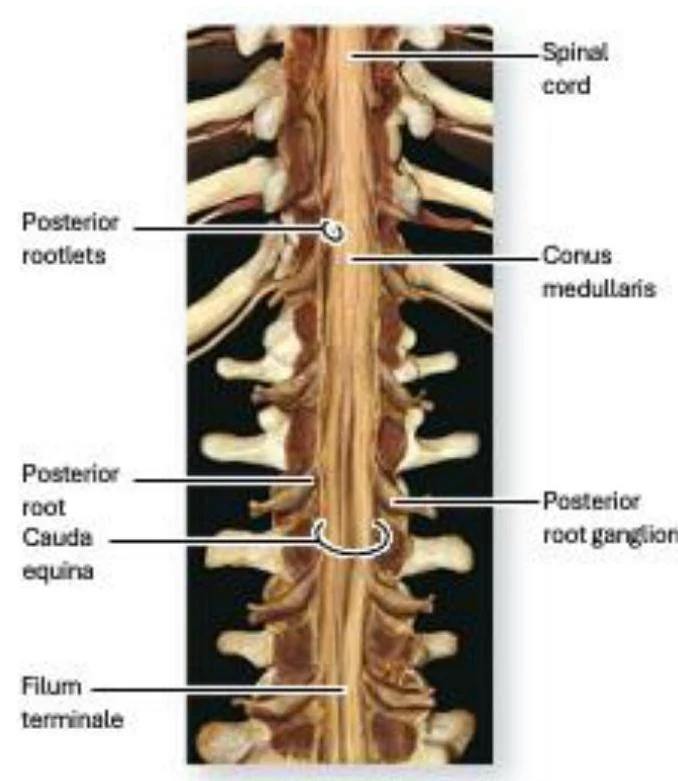
Because the spinal cord ends at L1, the lower portions of the vertebral canal contain a collection of nerve roots rather than the solid cord itself. The tapering, inferior end of the spinal cord is known as the conus medullaris.
Extending from the conus medullaris is a bundle of axons that resembles a horse's tail, called the cauda equina. To provide stability, a thin strand of pia mater called the filum terminale extends from the conus medullaris to attach it to the coccyx.
An illustration of the human spinal cord, showing the spinal cord, conus medullaris, cauda equina, posterior rootlets, posterior root ganglion, and filum terminale.
Spinal Meninges and Protection
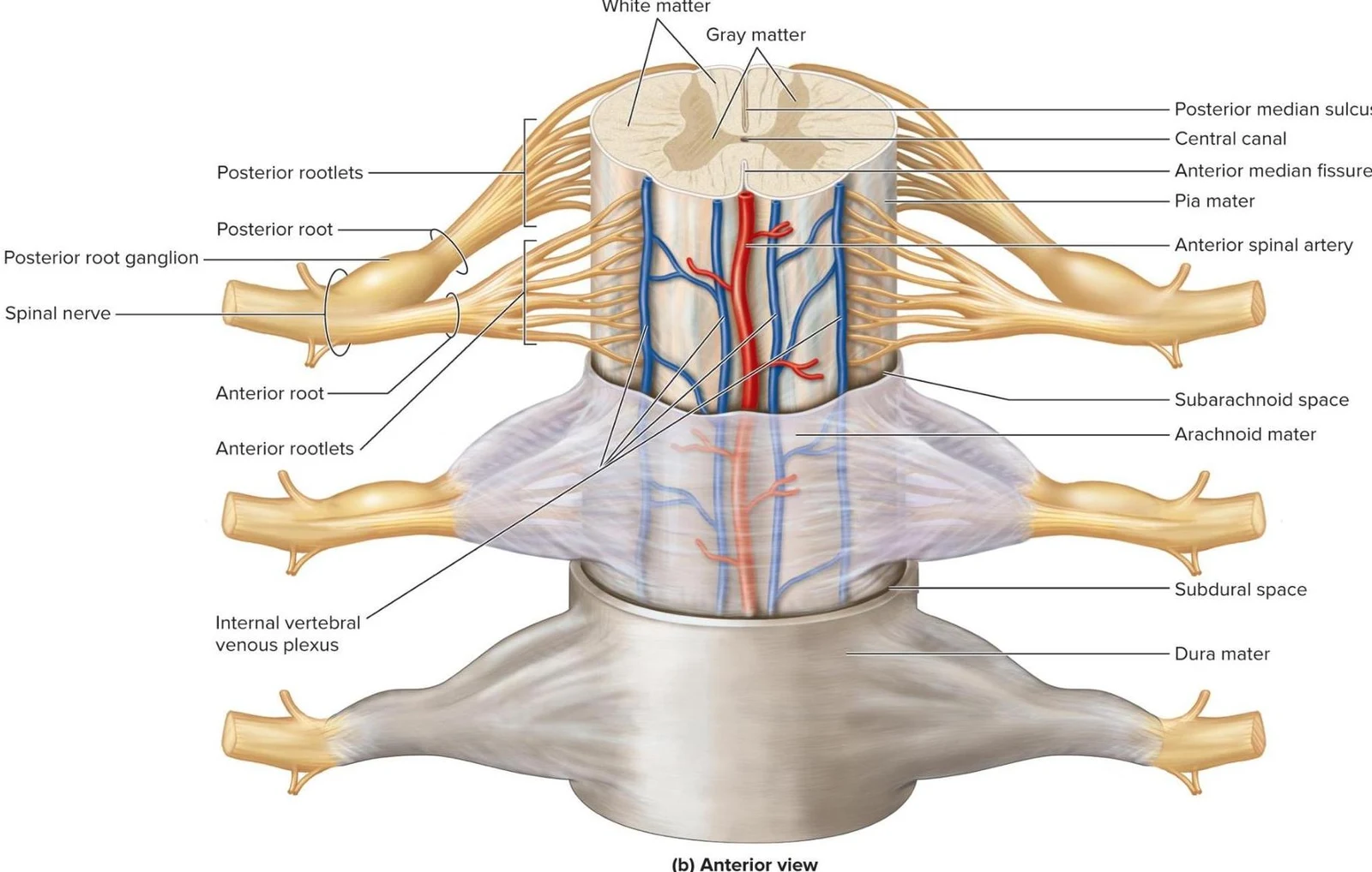
The spinal cord is protected by three layers of membranes called meninges, which are continuous with the cranial meninges:
Dura mater: The outermost, single-layered membrane that stabilizes the cord and fuses with the connective tissue of spinal nerves.
Arachnoid mater: The middle layer.
Pia mater: The innermost, delicate layer of elastic and collagen fibers that adheres directly to the spinal cord.
Between these layers lie critical spaces. The epidural space sits between the dura mater and the periosteum of the vertebra; this fat-filled space is where epidural anesthesia is administered. The subdural space is a narrow potential space between the dura and arachnoid mater. Finally, the subarachnoid space is a real space filled with cerebrospinal fluid (CSF).
Clinical Note: Lumbar Puncture
To sample CSF, a needle must pass through the skin, muscles, ligamentum flavum, epidural space, dura, and arachnoid to reach the subarachnoid space. To avoid piercing the spinal cord, this is typically performed at the L3-L4 or L4-L5 vertebrae, as the spinal cord is absent between L2 and S2.
To help anchor the cord laterally, the pia mater forms paired, triangular extensions called denticulate ligaments that attach the cord to the dura mater.
An anterior view of the spinal cord, illustrating its cross-section with white and gray matter, central canal, and surrounding meninges (pia mater, arachnoid mater, dura mater). Spinal nerves, roots, rootlets, and the internal vertebral venous plexus are also depicted.
Internal Structure: Gray and White Matter
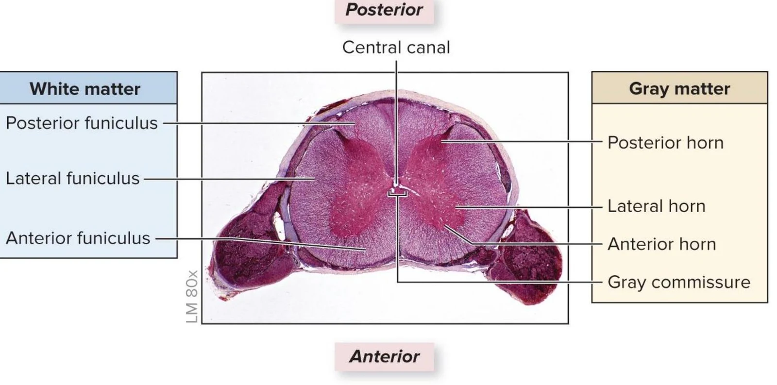
A cross-section of the spinal cord reveals a distinct organization of tissue: an inner region of gray matter and an outer region of white matter.
Gray Matter and the Horns
The gray matter contains neuron cell bodies, dendrites, unmyelinated axons, and glial cells. It is organized into "horns":
Posterior horns: Contain axons of sensory neurons and cell bodies of interneurons.
Lateral horns: Contain cell bodies of autonomic motor neurons; these are only found in the T1–L2 segments.
Anterior horns: House the cell bodies (somas) of somatic motor neurons.
The gray matter is bisected by the gray commissure, which contains unmyelinated axons for communication between the left and right sides, and houses the narrow central canal.
White Matter and Pathways
The white matter consists of myelinated axons organized into columns called funiculi (singular: funiculus): the posterior, lateral, and anterior funiculi. Within these funiculi, axons are organized into functional units called tracts.
Ascending (Sensory) pathways: Carry information from the body to the brain.
Descending (Motor) pathways: Transmit information from the brain to muscles or glands.
This is a cross-section of the spinal cord, illustrating the distinct regions of white matter and gray matter. The white matter is divided into the posterior, lateral, and anterior funiculi, while the gray matter is characterized by the posterior horn, lateral horn, anterior horn, and gray commissure. The central canal is also visible in the center.
Spinal Nerves and Plexuses
The spinal cord is associated with 31 pairs of spinal nerves: 8 cervical, 12 thoracic, 5 lumbar, 5 sacral, and 1 coccygeal.
After exiting the intervertebral foramen, a spinal nerve splits into rami:
Posterior ramus: Innervates the deep muscles and skin of the back.
Anterior ramus: Innervates the anterior/lateral trunk and the limbs.
Many anterior rami interweave to form nerve plexuses. Major plexuses include the cervical, brachial, lumbar, and sacral plexuses.
The Cervical Plexus
Formed by the anterior rami of C1–C4, the cervical plexus innervates the neck, skin of the head/shoulders, and anterior neck muscles. A vital branch is the phrenic nerve (primarily C4), which travels to the diaphragm to control breathing.
Exam Tip: Cutaneous Branches of the Cervical Plexus
To remember the cutaneous branches, use the mnemonic "Get LoST":
Great auricular (C2-C3)
Lesser occipital (C2)
Supraclavicular (C3-C4)
Transverse cervical (C2-C3)
Reflex Arcs
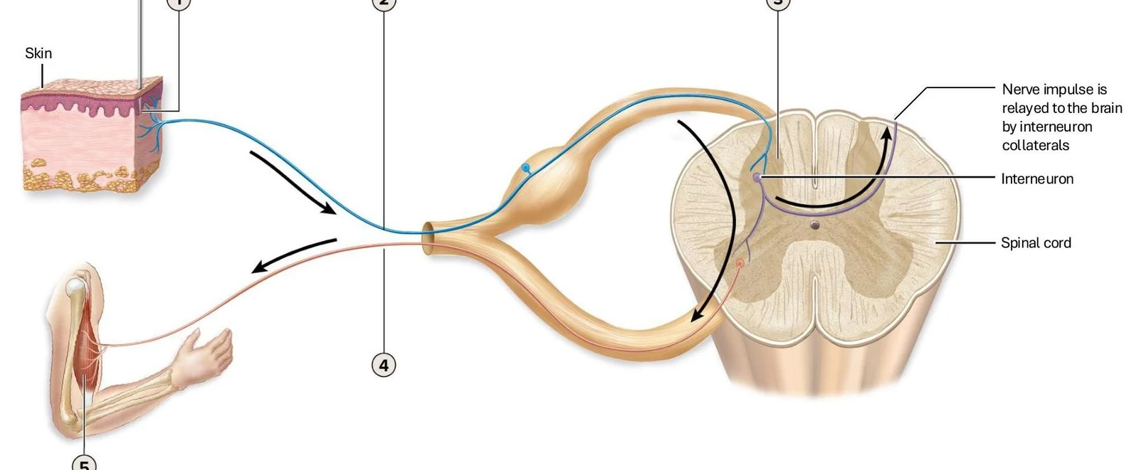
Reflexes are rapid, involuntary, and preprogrammed responses to a stimulus. A reflex arc is the neural circuit that makes this possible, consisting of five steps:
Stimulus activates a sensory receptor.
Impulse travels via a sensory neuron to the CNS.
Interneurons process the information.
Motor neurons send impulses to an effector.
The effector (muscle or gland) responds.
Reflexes can be categorized by their complexity and side of the body:
Monosynaptic: Only one synapse; the sensory axon synapses directly onto a motor neuron (e.g., the stretch reflex or knee-jerk reflex).
Polysynaptic: Involves one or more interneurons (e.g., the withdrawal reflex or the Golgi tendon reflex).
Ipsilateral: Receptor and effector are on the same side.
Contralateral: The impulse crosses the spinal cord to the opposite side.
Diagram illustrating a reflex arc, showing sensory input from the skin, processing in the spinal cord via an interneuron, and motor output to a muscle.
The Sympathetic Division
The sympathetic division (thoracolumbar division) is driven by preganglionic neuron cell bodies located in the lateral horns of T1 through L2.
Axons exit via the anterior roots and travel through rami communicantes to reach the sympathetic trunk (a chain of ganglia lateral to the vertebral column).
White rami communicantes: Carry myelinated preganglionic axons.
From the sympathetic trunk, pathways can follow several routes:
Spinal nerve pathway: Axons exit via gray rami to join spinal nerves.
Splanchnic nerve pathway: Preganglionic axons pass through the trunk without synapsing and terminate in prevertebral ganglia (like the celiac or mesenteric ganglia) near the abdominal organs.
Adrenal medulla pathway: Preganglionic axons travel directly to the adrenal gland, triggering the release of epinephrine and norepinephrine into the blood.
Keep reading for free.
Create an account to finish this reading and make it part of your study routine.