Anatomy and Physiology of the Spinal Cord and Nerves
A comprehensive overview of the spinal cord's structure, including its external and sectional anatomy, protective meninges, and the network of spinal nerves and plexuses. It also details clinical conditions and injuries related to the spinal cord.
The spinal cord is a vital neurological structure that serves as the primary communication link between the brain and the rest of the body. It functions as a high-speed pathway for sensory and motor impulses and acts as a central hub for spinal reflexes. Understanding its anatomy requires looking at its external structure, its protective coverings, its internal organization, and the complex network of nerves that emerge from it.
External Anatomy of the Spinal Cord
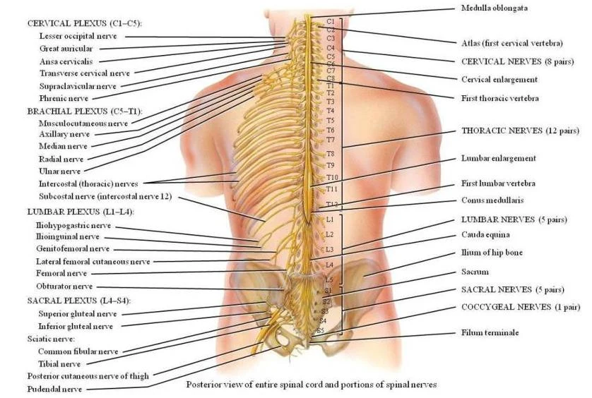
The spinal cord is approximately 16 to 18 inches long and extends from the foramen magnum of the skull down to the level of the $L1$ vertebra. It is divided into five distinct regions: cervical, thoracic, lumbar, sacral, and coccygeal.
As the spinal cord descends, it eventually tapers to a point known as the conus medullaris. Because the spinal cord is shorter than the vertebral column, several nerve roots must extend further down to reach their respective exit points. This collection of axons is called the cauda equina (Latin for "horse's tail"). To provide stability, a thin strand of pia mater called the filum terminale extends from the conus medullaris to anchor the cord to the coccyx.
To accommodate the complex innervation required for movement, the spinal cord features two significant enlargements:
Cervical enlargement (C4–T1): Provides innervation to the upper limbs.
Lumbosacral (Lumbar) enlargement (T9–T12): Provides innervation to the lower limbs.
Posterior view of the human spinal cord and associated nerves, showing the cervical, thoracic, lumbar, and sacral plexuses.Diagram illustrating the posterior view of the human spinal cord and its associated nerve plexuses, including the cervical, brachial, lumbar, sacral, and coccygeal nerves.
The external surface of the cord is marked by two longitudinal depressions: the deep, wide anterior median fissure on the ventral side and the shallow, narrow posterior median sulcus on the dorsal side.
The Meninges and Protection
The spinal cord is protected by three layers of connective tissue called meninges, which are continuous with the cranial meninges.
Dura mater: The outermost, tough layer that stabilizes the cord and fuses with the connective tissue surrounding the spinal nerves. The space between the dura mater and the periosteum of the vertebra is known as the epidural space.
Arachnoid mater: The middle layer, characterized by a web-like network of fibers called arachnoid trabeculae. Between the dura and the arachnoid lies a potential space called the subdural space, which contains interstitial fluid.
Pia mater: The innermost layer that adheres directly to the surface of the spinal cord. It uses denticulate ligaments to suspend and anchor the spinal cord laterally to the dura mater.
The subarachnoid space, located between the arachnoid mater and the pia mater, is filled with cerebrospinal fluid (CSF).
Clinical Correlation: Lumbar Puncture
A lumbar puncture (spinal tap) involves inserting a long, hollow needle into the subarachnoid space after administering local anesthesia. This procedure allows clinicians to withdraw CSF for diagnosis, introduce antibiotics or chemotherapy, measure CSF pressure, or evaluate diseases like meningitis (inflammation of the meninges caused by infection).
Sectional Anatomy: Gray and White Matter
When viewed in cross-section, the spinal cord reveals a distinct organization of tissue:
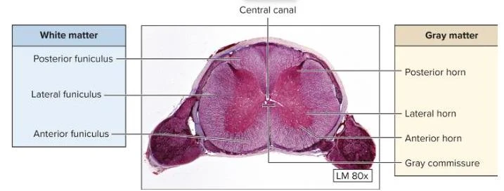
Cross-section of the spinal cord showing white matter and gray matter with labeled regions including posterior funiculus, lateral funiculus, anterior funiculus, posterior horn, lateral horn, anterior horn, gray commissure, and central canal.
Gray Matter
The central region consists of gray matter, which contains neuron cell bodies, dendrites, unmyelinated axons, and glial cells. It is organized into:
Anterior, posterior, and lateral horns.
Gray commissure: A bridge of unmyelinated axons that facilitates communication between the right and left sides of the cord.
Central canal: A small opening in the center of the gray commissure that runs the length of the cord.
White Matter
Surrounding the gray matter is the white matter, which consists of bundles of myelinated axons. These bundles are organized into columns known as funiculi (anterior, posterior, and lateral), which serve as sensory tracts.
Spinal Nerves and Plexuses
There are 31 pairs of spinal nerves emerging from the cord: 8 cervical, 12 thoracic, 5 lumbar, 5 sacral, and 1 coccygeal. Each nerve is formed by the merger of two roots:
Anterior (ventral) root: Carries motor axons to effectors like muscles and glands.
Posterior (dorsal) root: Carries sensory axons to the spinal cord.
After exiting the intervertebral foramen, the spinal nerve divides into rami:
Posterior (dorsal) ramus: Innervates the deep muscles and skin of the back.
Anterior (ventral) ramus: Innervates the limbs and the skin of the lateral and anterior trunk.
Rami communicantes: Connect the spinal cord to the sympathetic ganglia.
Nerve Plexuses
The anterior rami interweave to form complex networks called nerve plexuses:
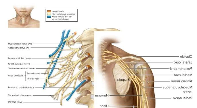
Cervical Plexus (C1–C4): Includes the phrenic nerve, which innervates the diaphragm.
Brachial Plexus (C5–T1): Innervates the upper limbs. Key nerves include the musculocutaneous (biceps), median and ulnar (forearm flexors), radial (extensors), and axillary (deltoid) nerves.
An illustration of the nerves of the neck and shoulder, including the cervical plexus and brachial plexus.
Lumbar Plexus (L1–L4): Includes the femoral nerve (hip flexors/knee extensors) and the obturator nerve (hip adductors).
Sacral Plexus (L4–S4): Includes the massive sciatic nerve, which divides into the tibial and common fibular nerves to innervate the posterior thigh, leg, and foot.
Dermatomes
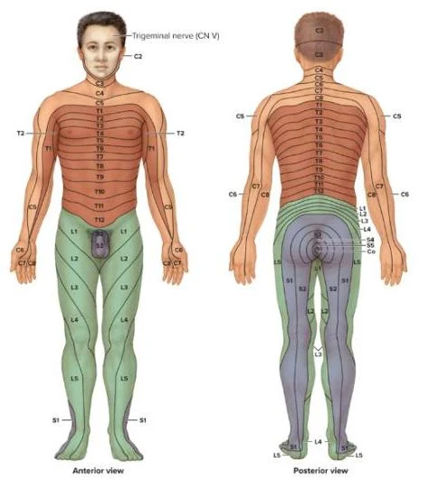
A dermatome is an area of skin innervated by a single spinal nerve. For example, T4 corresponds to the nipple level, T10 to the umbilicus, and S1 to the plantar region of the foot.
Anterior and posterior views of the human body illustrating dermatomes, which are areas of skin supplied by a single spinal nerve.
Clinical Conditions and Injuries
Damage to the spinal cord or its nerves can result in various neurological deficits:
Paralysis Types:
Monoplegia: One limb.
Paraplegia: Both lower limbs.
Quadriplegia: All four limbs.
Hemiplegia: One upper and one lower limb on the same side.
Specific Nerve Injuries:
Wrist Drop: Injury to the radial nerve, preventing extension of the wrist.
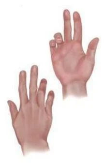
Clawhand: Ulnar nerve palsy resulting in hyperextension of metacarpophalangeal joints.
Foot Drop: Injury to the common fibular nerve causing plantar flexion.
Sciatica: Pain extending from the gluteal region down the leg, often caused by a herniated disc.
Exam Tip: Spinal Shock
If a question asks about the immediate response to a spinal cord injury, remember spinal shock, which is characterized by temporal areflexia (a temporary loss of reflex function).
Illustration depicting wrist drop, a condition characterized by the inability to extend the wrist and fingers.Illustration of two hands showing different finger positions.
Keep reading for free.
Create an account to finish this reading and make it part of your study routine.