Anatomy and Physiology of the Small and Large Intestines
This content details the anatomy and physiological functions of the small and large intestines, including their roles in digestion, nutrient absorption, and waste processing. It covers specialized structures, chemical digestion, and absorption mechanisms.
The digestive process is a complex sequence of mechanical and chemical events designed to transform food into absorbable subunits. While the stomach initiates the breakdown of large proteins, the true heavy lifting of digestion and nutrient absorption occurs within the intestines. The small intestine serves as the primary site for chemical digestion and nearly all nutrient absorption, while the large intestine focuses on water reclamation and waste preparation.
Anatomy of the Small Intestine
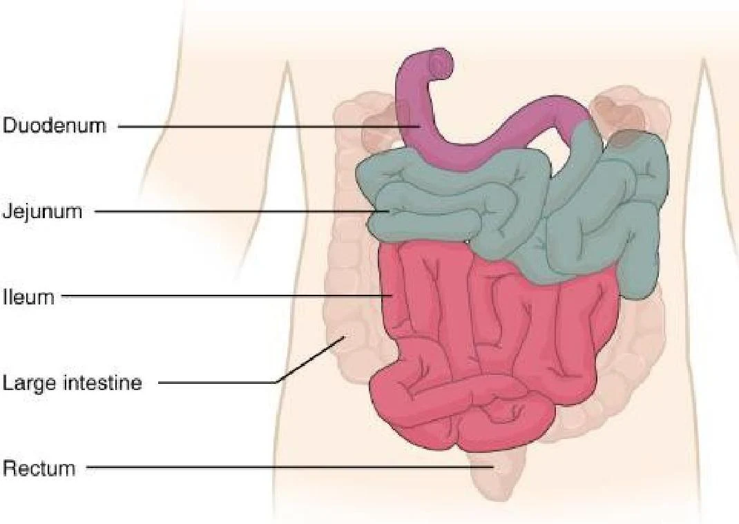
The small intestine is the longest portion of the gastrointestinal (GI) tract. It is divided into three distinct anatomical regions:
Duodenum: The first section, which connects to the stomach via the pyloric sphincter. It receives chyme, bile, and pancreatic enzymes.
Jejunum: The middle section where much of the nutrient absorption occurs.
Ileum: The final and longest section of the small intestine, which leads into the large intestine through the ileocecal valve.
Diagram of the human digestive system, highlighting the duodenum, jejunum, ileum, large intestine, and rectum.
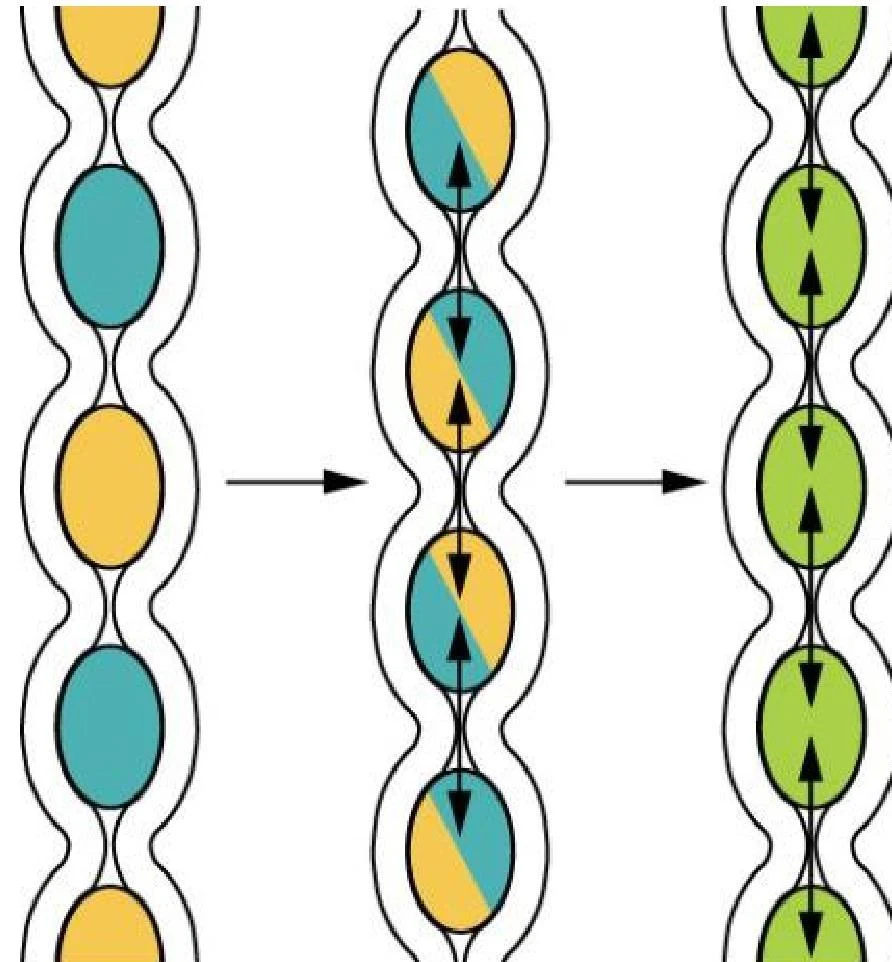
To maximize efficiency, the small intestine utilizes segmentation. This mechanical process involves the contraction of intestinal muscles to separate chyme and then push it back together, mixing the contents thoroughly and providing sufficient time for enzymes to work and nutrients to be absorbed.
Diagram illustrating a process of change over time, possibly related to cell division or particle transformation, shown in three stages.
Specialized Structures for Absorption
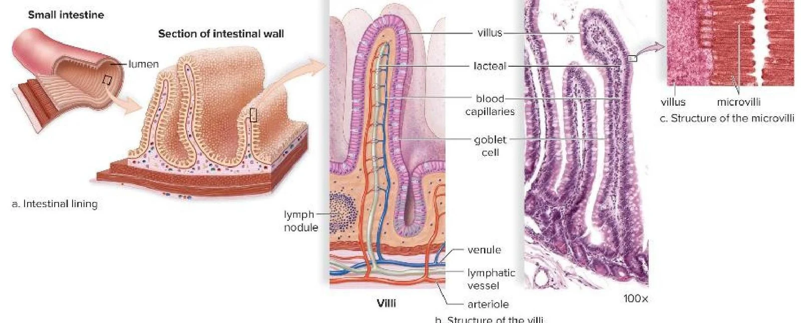
The small intestine is uniquely adapted to absorb nutrients through a massive increase in surface area. This is achieved through three hierarchical levels of folding:
Circular Folds: Large inward folds of the intestinal wall.
Villi: Fingerlike projections that extend from the mucosa.
Microvilli: Microscopic projections on the surface of the individual cells covering the villi.
Diagrams illustrating the structure of the small intestine, including its lining, villi, and microvilli.
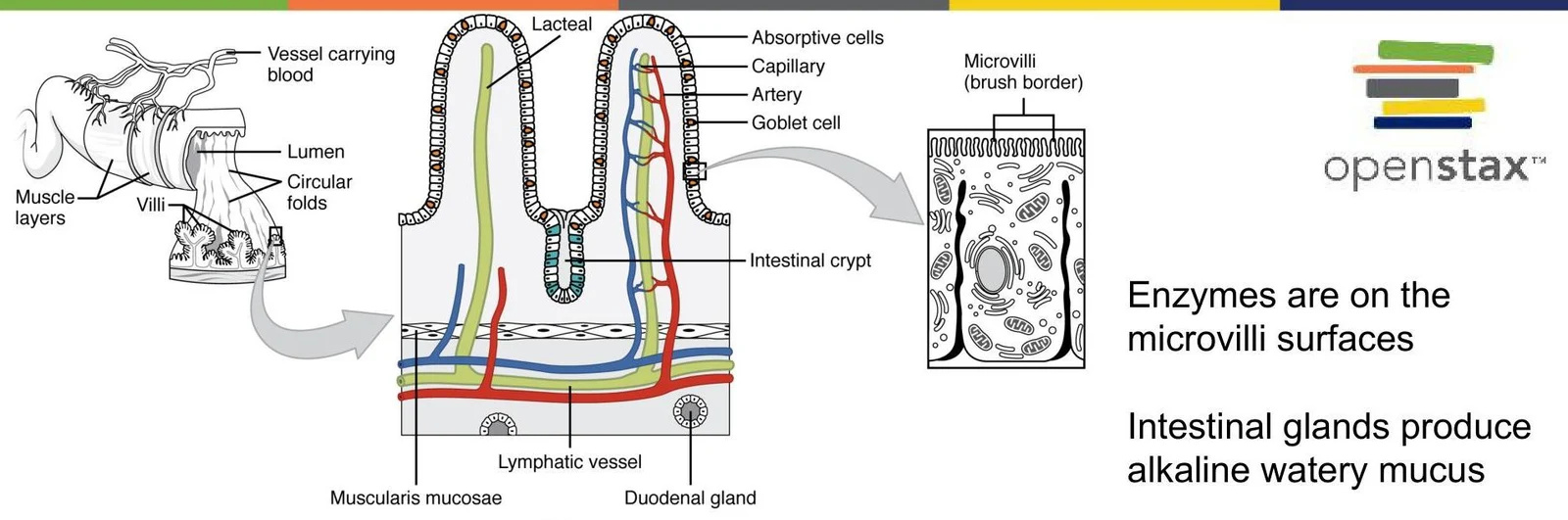
The presence of microvilli creates what is known as the brush border. This border is not just structural; it contains "brush border enzymes" that perform the final stages of chemical digestion.
Diagram illustrating the structure of the small intestine, including villi, microvilli, and associated blood and lymphatic vessels, along with the functions of intestinal glands and enzymes.
Chemical Digestion and Nutrient Absorption
Chemical digestion involves the hydrolysis of macromolecules into subunits small enough to cross plasma membranes. This process is driven by various digestive enzymes produced by the salivary glands, stomach, pancreas, and the small intestine itself.
Breakdown of Macromolecules
The specific enzymes required depend on the type of nutrient being processed:
Carbohydrates: Digestion begins in the mouth with salivary amylase and is completed in the small intestine by pancreatic amylase and brush border enzymes like maltase and lactase, resulting in monosaccharides (glucose, galactose, and fructose).
Proteins: Digestion starts in the stomach with pepsin and continues in the small intestine with pancreatic trypsin and intestinal peptidases, breaking proteins down into single amino acids or small peptides.
Nucleic Acids: Broken down into nucleotides by pancreatic nucleases and subsequently into pentose sugars, phosphates, and nitrogenous bases by nucleosidases.
Fats (Lipids): Virtually all fat digestion occurs in the small intestine. Pancreatic lipase breaks triglycerides into glycerol and free fatty acids.
The Absorption Process
Once broken down, nutrients must be transported into the body. The method of transport varies by substance:
Water-Soluble Nutrients: Monosaccharides, amino acids, and electrolytes are absorbed into the blood capillaries within the villi via mechanisms such as active transport, facilitated diffusion, or cotransport.
Lipids: Because fats are not water-soluble, they undergo a unique process. After being broken down, they are reassembled into lipoprotein droplets called chylomicrons. These chylomicrons enter a specialized lymphatic capillary called a lacteal rather than the blood directly.
Exam Tip: Remember the distinction between transport routes! Water-soluble nutrients (sugars/amino acids) go to the blood, while fats (via chylomicrons) go to the lymph (lacteals).
Anatomy of the Large Intestine
The large intestine is the final segment of the GI tract. Unlike the small intestine, it lacks folds and villi. Its primary functions are to absorb salts and water, synthesize certain vitamins, and form and eliminate feces.
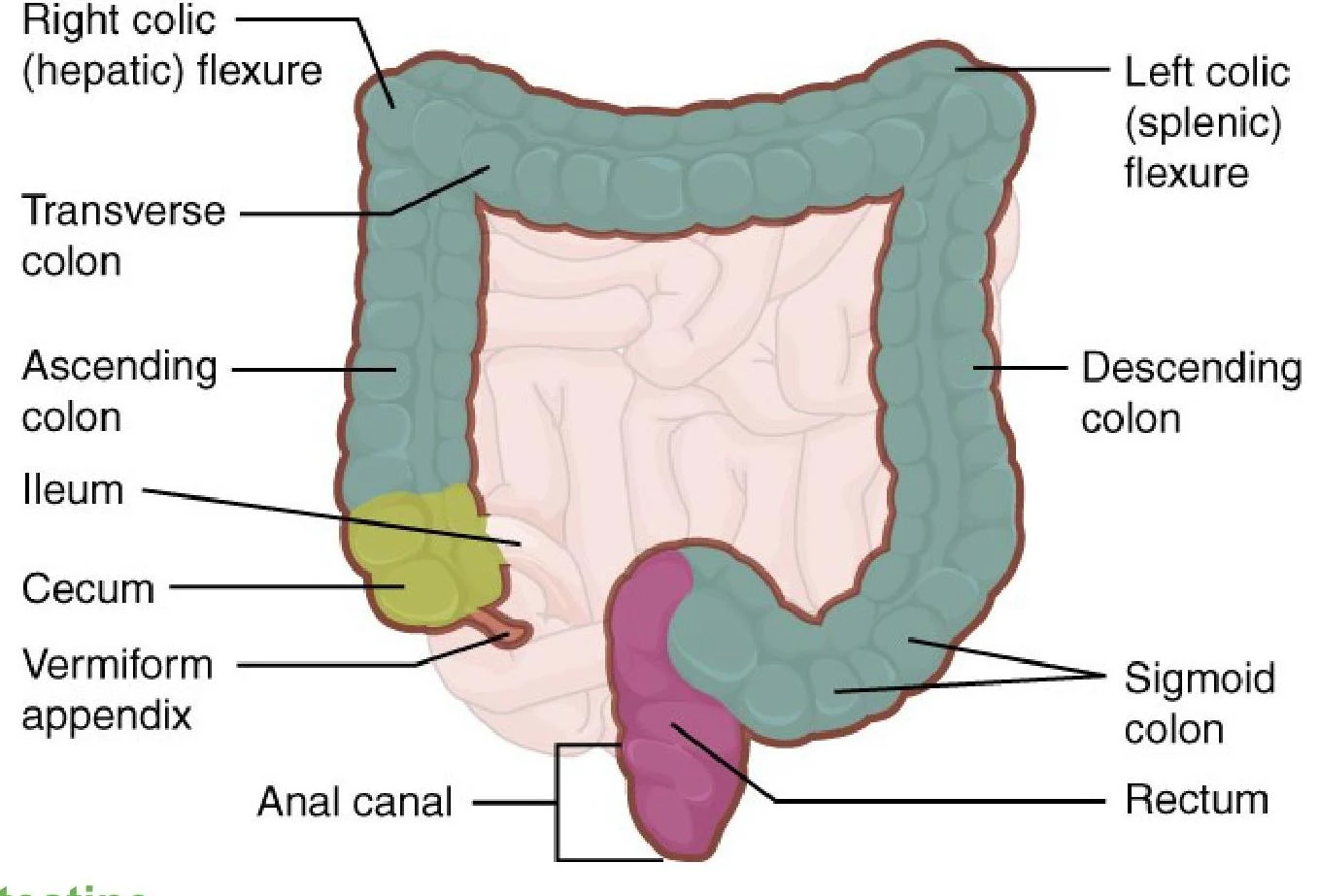
The large intestine is divided into the cecum, colon (ascending, transverse, and descending), rectum, and anus.
Diagram of the human large intestine, labeling the cecum, appendix, colon (ascending, transverse, descending, sigmoid), rectum, and anal canal, along with the hepatic and splenic flexures.
Unique Structural Features
The large intestine possesses several specialized features:
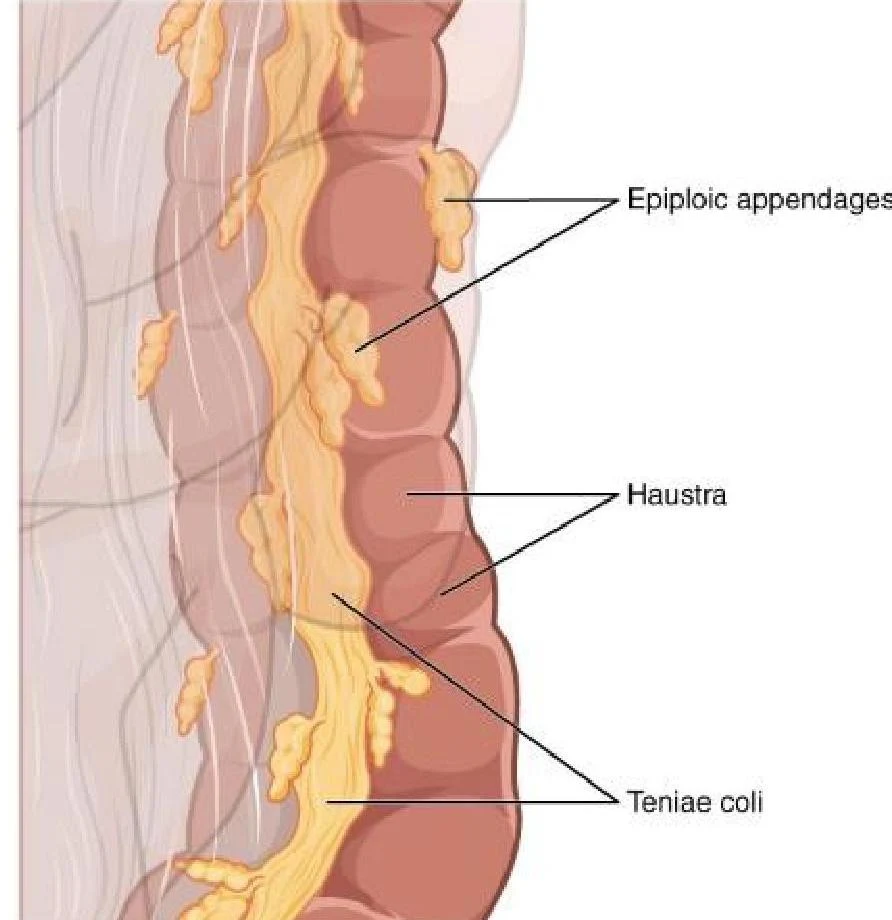
Teniae Coli: Three bands of smooth muscle that run along the longitudinal layer of the muscularis.
Haustra: Pouches formed by the contractions of the teniae coli.
Epiploic Appendages: Small, fat-filled sacs of visceral peritoneum.
An illustration of the colon showing epiploic appendages, haustra, and teniae coli.
Movement in the large intestine is generally slower than in the small intestine, characterized by haustral contractions and occasional "mass movements" that often follow meals. The final product, feces, consists of approximately 75% water and 25% undigested materials, including bacteria and electrolytes.
Keep reading for free.
Create an account to finish this reading and make it part of your study routine.