


The respiratory system is a complex network of organs and structures designed to facilitate pulmonary ventilation and the vital process of gas exchange. By moving air in and out of the body, the system ensures that oxygen reaches the bloodstream and that carbon dioxide, a metabolic waste product, is expelled. Understanding this system requires a dual focus on its physical architecture—from the protective bony cage of the thorax to the microscopic alveoli—and the mechanical physiology that drives the breath.
Anatomic Structures of the Thorax
The thorax is the anatomical region extending from the base of the neck down to the diaphragm. It serves as a protective housing for several vital organs, including the lungs, the liver, and major blood vessels.
The thorax is organized into two primary components: the thoracic cage and the thoracic cavity. The thoracic cage acts as a structural shield, composed of the sternum, the ribs, and various cartilages. This cage is further encased by layers of muscle, skin, and fasciae to provide both protection and flexibility.
Bony Landmarks and Clinical Reference Lines
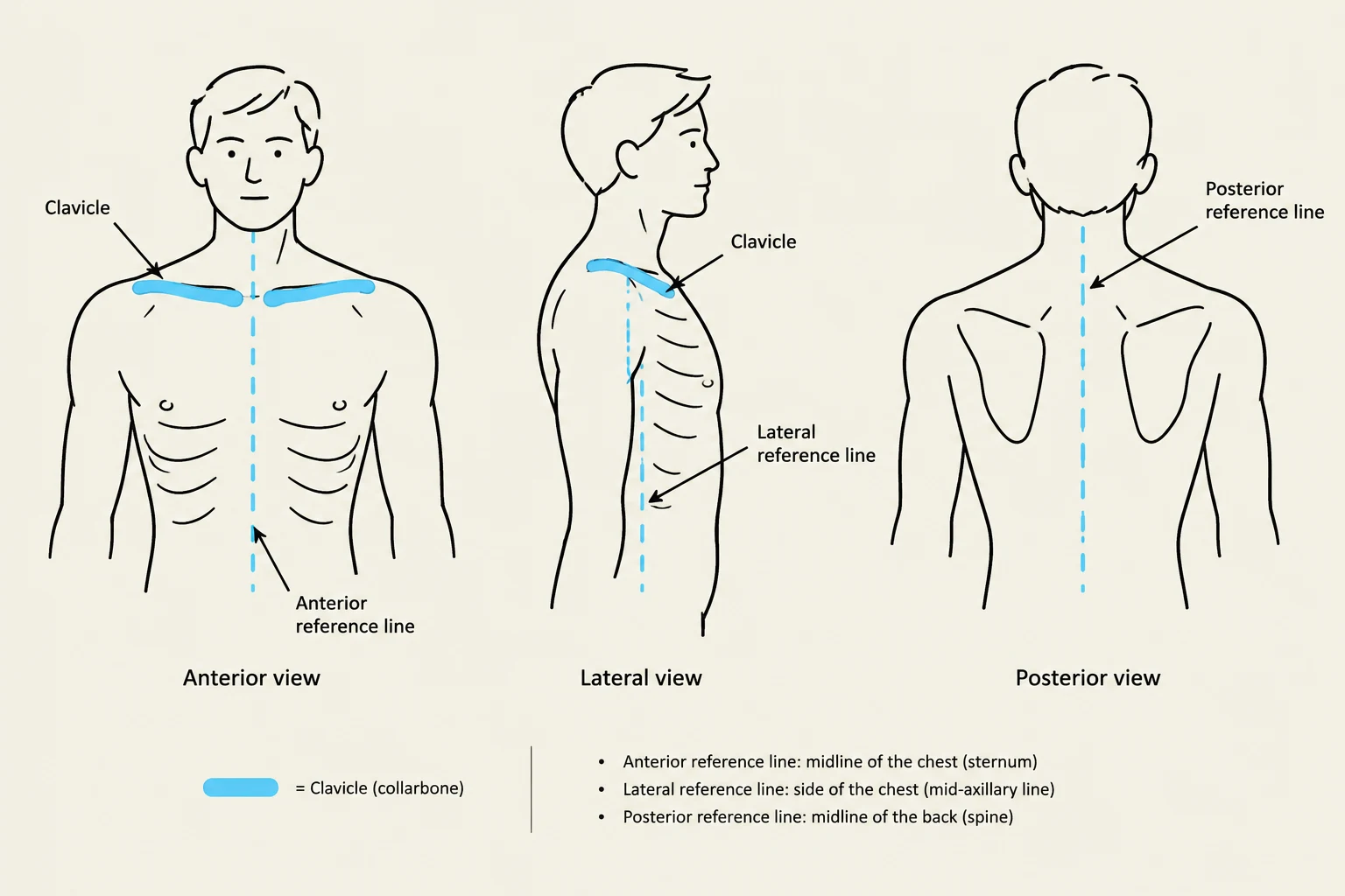
For clinicians, the ability to precisely locate anatomical structures is essential for physical assessment. To achieve this, clinicians utilize anterior, lateral, and posterior reference lines.
- Anterior Chest Wall: Assessment focuses on the clavicles, the sternum, the ribs, and the costal cartilages.
- Posterior Chest Wall: Assessment focuses on the vertebral column, the posterior aspects of the ribs, and the scapula.
- Lateral Chest Wall: Provides additional access points for evaluating the lung fields.

Keep reading for free.
Create an account to finish this reading and make it part of your study routine.
Continue reading freeAlready have an account? Log in
Updated