Anatomy and Physiology of the Pharynx and Esophagus
An overview of the pharynx and esophagus, detailing their anatomical divisions, muscular structures, and the physiological processes of swallowing and peristalsis.
The pharynx and esophagus are critical components of both the respiratory and digestive systems. While they serve different primary purposes—breathing and swallowing—they share a common pathway, necessitating highly coordinated muscular movements to ensure that food reaches the stomach without entering the airway. This article explores the anatomical divisions, the complex muscular arrangements of the pharynx, and the peristaltic mechanisms of the esophagus.
Functions of the Pharynx
The pharynx is a muscular tube that serves three primary roles:
Common Pathway: It provides a shared route for both food intake (digestive) and breathing (respiratory).
Swallowing Mechanism: It undergoes rapid contractions to clear food boluses from the pharyngeal cavity and move them into the esophagus.
Speech Resonance: It acts as a resonating chamber, which is essential for the production of various speech sounds.
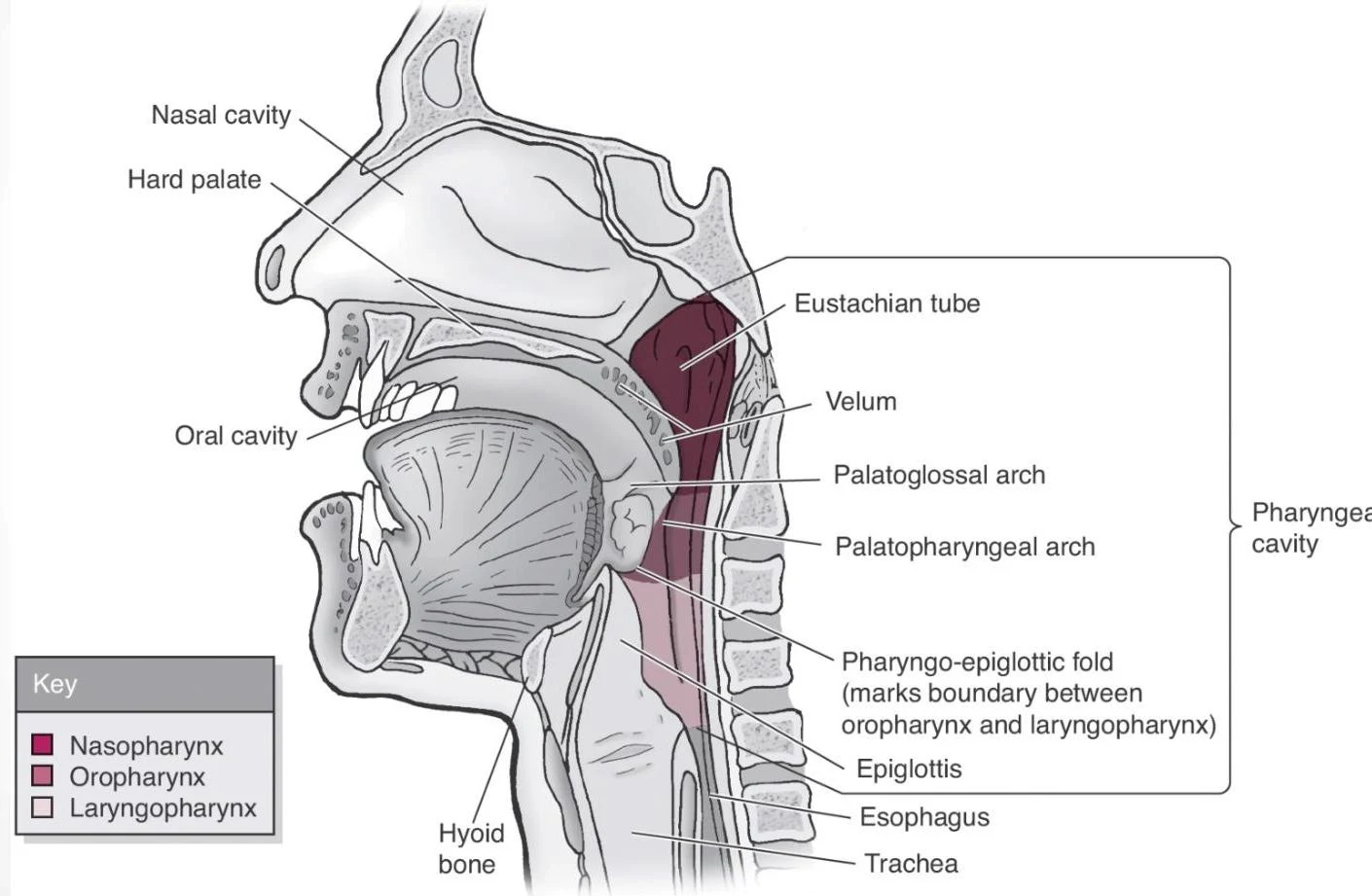
This diagram illustrates a sagittal view of the human pharynx, highlighting key anatomical structures such as the nasal cavity, oral cavity, Eustachian tube, epiglottis, esophagus, and trachea. The key indicates the divisions of the pharynx: nasopharynx, oropharynx, and laryngopharynx.
Anatomical Divisions of the Pharynx
The pharynx is divided into three distinct anatomical regions based on their location:
Nasopharynx: The uppermost portion, located behind the nasal cavity.
Oropharynx: The middle portion, located behind the oral cavity.
Laryngopharynx: The lowermost portion, located above the esophagus and behind the larynx.
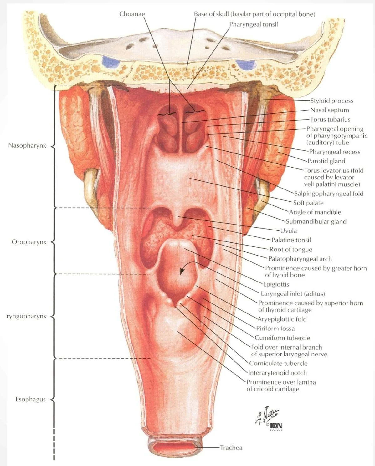
Anatomical illustration of the human pharynx and upper airway, detailing structures such as the nasopharynx, oropharynx, laryngopharynx, esophagus, and trachea, with labels for various anatomical landmarks.
Muscles of the Pharynx
The muscles of the pharynx are categorized into two functional groups: the constrictors, which squeeze the bolus downward, and the elevators, which lift the pharynx to facilitate swallowing.
The Pharyngeal Constrictors
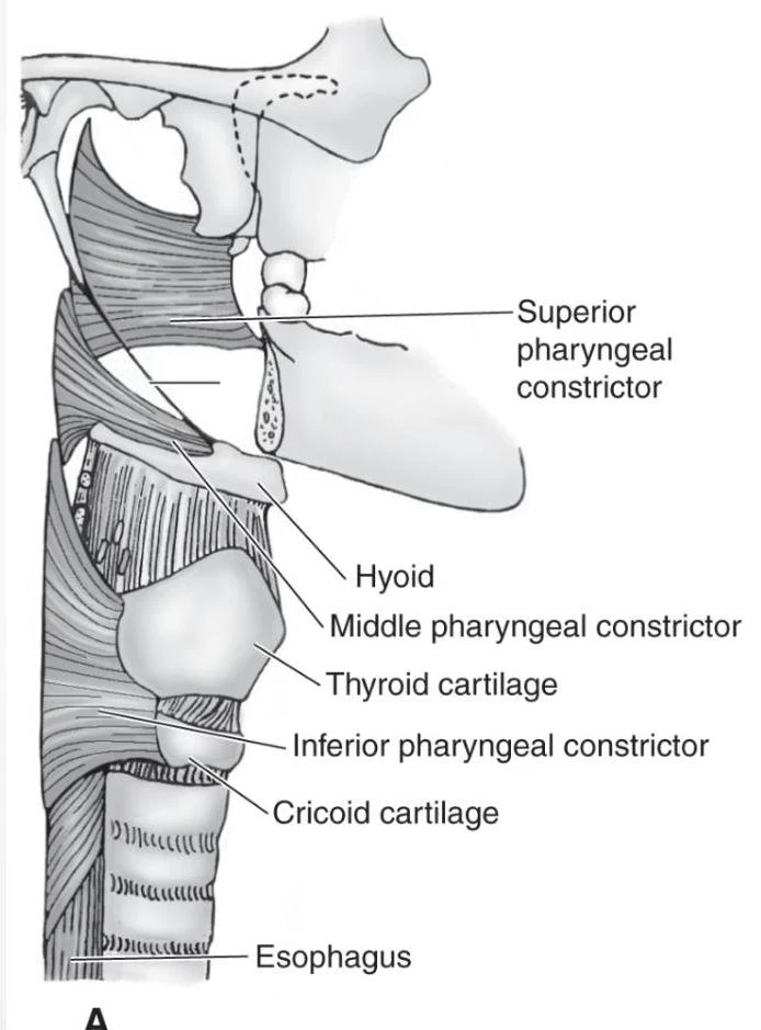
There are three constrictor muscles organized like "stacked cups". Most of these muscles are open anteriorly and connect to the posterior median pharyngeal raphe, with the exception of the lower fibers of the inferior constrictor, which form a complete ring.
Superior Constrictor ("1st cup"): This is the weakest of the three constrictors. It originates from the medial pterygoid plate and inserts into the posterior median pharyngeal raphe and the occipital bone. Its primary actions are pharyngeal constriction during swallowing and assisting in velopharyngeal closure. It is innervated by the Accessory nerve (CN XI).
Middle Constrictor ("2nd cup"): A fan-shaped muscle that originates from the stylohyoid and hyoid bones, inserting into the posterior median pharyngeal raphe. It is also innervated by the Accessory nerve (CN XI).
Inferior Constrictor ("3rd cup"): This is the thickest and strongest constrictor and is divided into two distinct parts:
Thyropharyngeus: Originates from the oblique line of the thyroid cartilage and inserts into the posterior median pharyngeal raphe.
Cricopharyngeus: A narrow band that originates and inserts on the cricoid cartilage. Its primary role is to keep the esophagus closed at rest by maintaining the Upper Esophageal Sphincter (UES). It is innervated by the pharyngeal branch, the external superior laryngeal nerve (eSLN), and the recurrent laryngeal nerve (RLN) of the Vagus nerve (CN X).
An anatomical illustration of the pharyngeal constrictor muscles, hyoid bone, thyroid cartilage, cricoid cartilage, and esophagus.
The Pharyngeal Elevators
Three additional muscles insert into the pharyngeal wall to elevate the pharynx during the swallowing process:
Salpingopharyngeus: A long, thin, paired muscle originating from the Eustachian tube and inserting into the lateral wall of the pharynx. It elevates and shortens the pharynx and helps keep the Eustachian tube closed at rest. It is innervated by CN XI.
Stylopharyngeus: A long, thin, paired muscle originating from the styloid process and inserting into the lateral wall of the pharynx. It elevates and shortens the pharynx and is innervated by the Glossopharyngeal nerve (CN IX).
Palatopharyngeus: This muscle forms the palatopharyngeal arch. It originates from the palatine raphe and inserts into the thyroid cartilage and the inferior portion of the pharynx. Its action is to lift the pharynx and larynx and direct the food bolus into the pharynx during swallowing. It is innervated by CN XI.
Anatomy and Physiology of the Esophagus
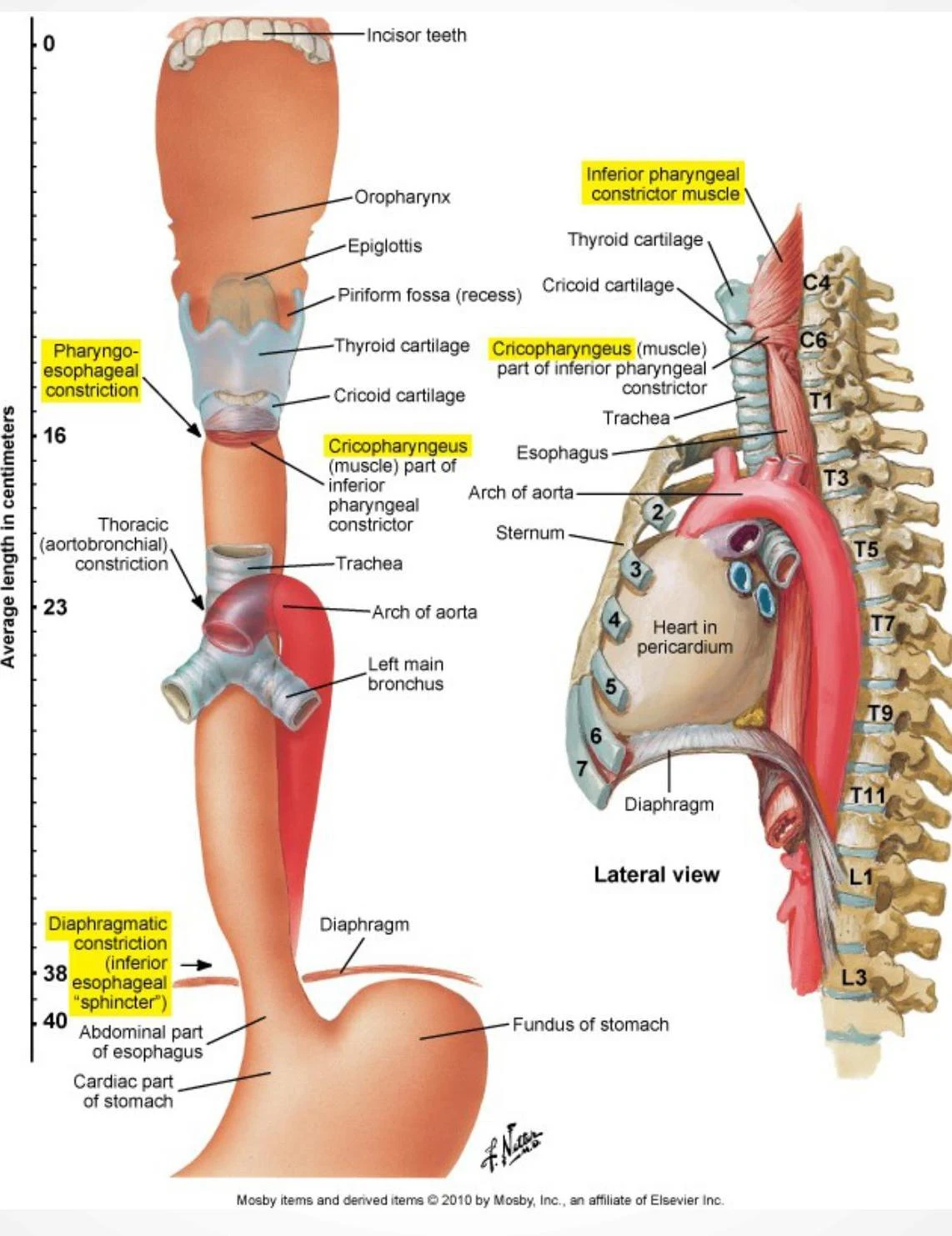
The esophagus is a collapsible, muscular tube approximately 25 cm in length. It is situated posterior to the trachea and anterior to the vertebral column, extending from the laryngopharynx down to the stomach, where it pierces the diaphragm through the esophageal hiatus.
Sphincters of the Esophagus
The esophagus is regulated by two functional sphincters that control the passage of food and prevent reflux:
Upper Esophageal Sphincter (UES): Located at the upper border of the esophagus, the UES is comprised of the cricopharyngeus muscle. It remains closed at rest due to the contraction of the cricopharyngeus; it opens only upon the relaxation of this muscle during swallowing.
Lower Esophageal Sphincter (LES): Located at the junction between the esophagus and the stomach at the level of the diaphragm. Unlike the UES, there is no specific sphincteric muscle present here; instead, the combined pressure of the esophageal muscles and the diaphragm maintains closure to prevent gastroesophageal reflux.
Peristalsis
The movement of food through the esophagus is achieved through two types of peristaltic waves:
Primary Peristalsis: This is the initial muscle contraction wave triggered by the arrival of a food bolus at the Upper Esophageal Sphincter. The contraction wave travels the entire length of the esophagus to move the bolus toward the stomach.
Secondary Peristalsis: If any food residue remains in the esophagus after the primary wave has passed, a second peristaltic wave is triggered locally to clear the remaining material.
Exam Tip: Remember that the Cricopharyngeus is part of the inferior pharyngeal constrictor, but it serves as the functional component of the Upper Esophageal Sphincter (UES).
Anatomical illustration of the human esophagus and surrounding structures, including the pharynx, larynx, trachea, aorta, heart, diaphragm, and stomach. The diagram highlights various constrictions and anatomical landmarks of the esophagus.
Keep reading for free.
Create an account to finish this reading and make it part of your study routine.