An overview of the human nervous system, including its organization into central and peripheral divisions, the functions of brain structures, cranial nerves, neurotransmission, and age-related changes.
The neurologic system is the body's most complex command center, responsible for coordinating everything from the most basic life-sustaining functions to the highest levels of human thought and emotion. To understand how the body responds to injury, disease, or aging, one must first understand the intricate organization of the nervous system, the chemical messengers that facilitate communication, and the specific functional divisions of the brain.
Organization of the Nervous System
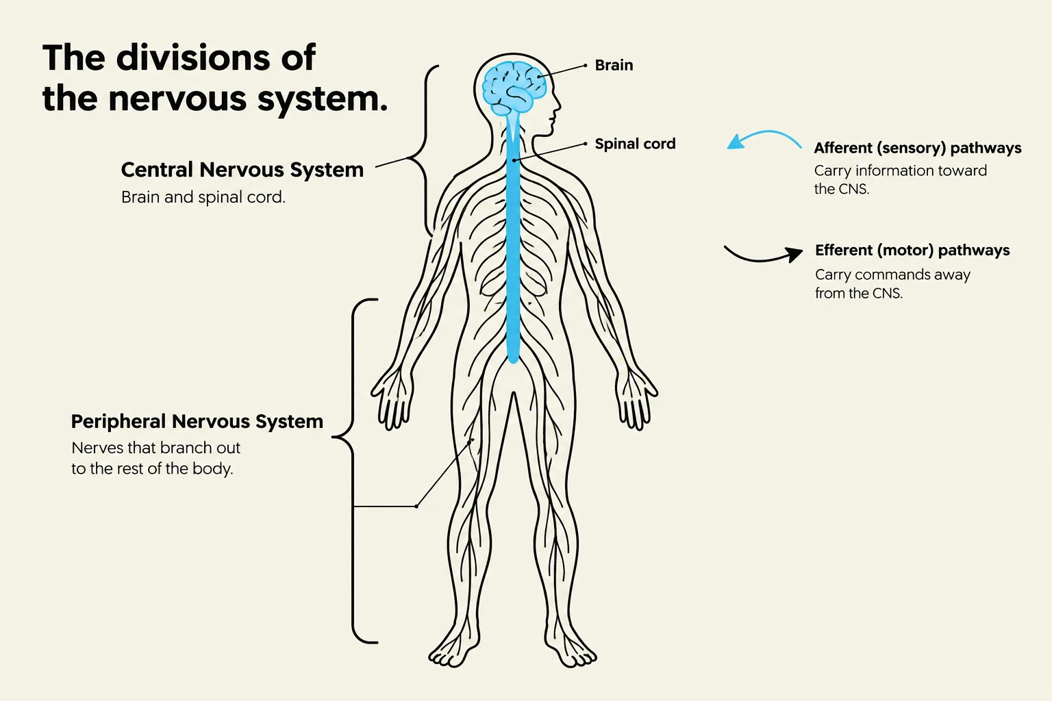
This diagram illustrates the divisions of the nervous system, including the Central Nervous System (brain and spinal cord) and the Peripheral Nervous System (nerves branching out to the body). It also depicts afferent (sensory) and efferent (motor) pathways.
The nervous system is broadly categorized into two main divisions: the Central Nervous System (CNS) and the Peripheral Nervous System (PNS).
The Central Nervous System (CNS)
The CNS serves as the primary processing center for the body. It consists of the brain and the spinal cord. Because the CNS is responsible for critical decision-making and vital life functions, its cells are incredibly specialized and fragile. A key clinical takeaway is that once neurons in the brain are destroyed, they cannot be replaced. Furthermore, these cells are highly sensitive to oxygen deprivation; if the brain experiences anoxia (a total lack of oxygen), irreversible damage can occur within just 4 to 6 minutes.
The Peripheral Nervous System (PNS)
The PNS acts as the communication relay between the CNS and the rest of the body. It is composed of cranial nerves and spinal nerves. Unlike the CNS, the PNS possesses a limited capacity for repair; specifically, Schwann cells in the PNS can allow for some nerve regeneration after injury.
The PNS is further organized by the direction of information flow:
Afferent Division: Carries sensory information from the body toward the CNS.
Efferent Division: Carries motor commands from the CNS out to the muscles and glands.
The PNS also contains the autonomic nervous system, which regulates involuntary functions through two opposing branches:
Sympathetic System: Prepares the body for "fight or flight" responses.
Parasympathetic System: Promotes "rest and digest" activities to maintain homeostasis.
Clinical Note: The Reflex Arc
A critical interaction between the CNS and PNS is the reflex arc. This is a rapid, involuntary pathway that allows the body to respond to stimuli (like pulling a hand away from a hot stove) before the brain even processes the sensation of pain.
Functional Divisions of the Brain
Diagram illustrating the major anatomical regions of the human brain, including the cerebrum, thalamus, hypothalamus, midbrain, pons, medulla oblongata, and cerebellum.
The brain is not a single uniform mass; it is divided into distinct regions, each with specialized responsibilities.
The Cerebrum
The cerebrum is the largest part of the brain and is responsible for higher-order functions, including thought, reasoning, memory, and voluntary movement.
The Cerebellum
Located beneath the cerebrum, the cerebellum is essential for coordinating movement, maintaining posture, and ensuring balance.
The Diencephalon
This central region contains two vital structures:
Thalamus: Acts as the sensory relay station, sorting and directing sensory input to the correct areas of the cerebrum.
Hypothalamus: The master regulator of homeostasis, controlling temperature, hunger, thirst, and the endocrine system.
The Brainstem
The brainstem connects the cerebrum to the spinal cord and controls the most fundamental life-sustaining functions (such as breathing and heart rate). It is composed of three parts:
Midbrain: Involved in visual and auditory reflexes.
Pons: Acts as a bridge between different parts of the brain and helps regulate breathing.
Medulla Oblongata: The most critical part for vital life functions, including cardiac and respiratory control.
Cranial Nerves
There are twelve pairs of cranial nerves (CN) that emerge directly from the brain to govern the sensory and motor functions of the head and neck.
Nerve Pair
Name
Primary Function
CN I
Olfactory
Smell
CN II
Optic
Vision
CN III, IV, VI
Oculomotor, Trochlear, Abducens
Eye movement
CN V
Trigeminal
Facial sensation and chewing
CN VII
Facial
Facial expression and taste
CN VIII
Acoustic (Vestibulocochlear)
Hearing and balance
CN IX
Glossopharyngeal
Taste and swallowing
CN X
Vagus
Autonomic functions (heart, lungs, etc.)
CN XI
Spinal Accessory
Shoulder and neck movement
CN XII
Hypoglossal
Tongue movement
Neurotransmission and Nerve Conduction
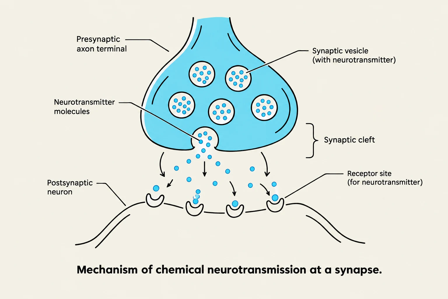
Diagram illustrating the mechanism of chemical neurotransmission at a synapse, showing the release of neurotransmitters from a presynaptic neuron and their binding to receptors on a postsynaptic neuron.
Communication within the nervous system relies on the conduction of electrical impulses and the release of chemical messengers known as neurotransmitters. When an impulse reaches the end of a neuron, neurotransmitters are released into the synapse to carry the signal to the next cell.
Several key neurotransmitters influence how these impulses are transmitted:
Acetylcholine: Crucial for muscle contraction and parasympathetic signals.
Dopamine & Norepinephrine: Involved in reward, motivation, and the sympathetic "fight or flight" response.
Epinephrine: Often released during stress to increase heart rate and energy.
Serotonin: Plays a major role in mood regulation and sleep.
GABA (Gamma-aminobutyric acid): The primary inhibitory neurotransmitter, which helps calm neural activity.
Glutamate (Glutamic acid): The primary excitatory neurotransmitter, essential for learning and memory.
Endorphins and Enkephalins: The body's natural opioids, responsible for pain suppression and pleasure.
Age-Related Changes
As the human body ages, the neurologic system undergoes significant physiological shifts. These changes can impact how an individual processes information and maintains physical stability.
Key changes include:
Structural Loss: A general loss of neurons and a decrease in overall blood flow to the brain.
Degeneration: In the PNS, the myelin sheath (the protective coating of nerves) and motor nerve fibers begin to degenerate.
Chemical Declines: Decreased secretion of neurotransmitters like norepinephrine and dopamine.
Functional Impairments:
Diminished or absent reflexes.
Smaller pupil size.
Impaired short-term memory.
Increased time required for cognitive processing, problem-solving, or learning new information.
Student Tip: Promoting Brain Health
While some age-related changes are inevitable, research suggests that keeping the brain active through mental stimulation can help promote continued intellectual function in healthy older adults.
Keep reading for free.
Create an account to finish this reading and make it part of your study routine.