Anatomy and Physiology of the Human Vascular System
An in-depth exploration of the human vascular system, detailing the structure and function of arteries, veins, and capillaries. Covers systemic and pulmonary circulation, major vessels of the head, neck, trunk, and extremities, and clinical applications.
The human vascular system is a complex network of vessels designed to transport blood throughout the body, ensuring that every tissue receives oxygen and nutrients while metabolic wastes are removed. This system is divided into two primary circuits: the systemic circulation, which extends to all body regions, and the pulmonary circulation, which facilitates gas exchange in the lungs. To understand how blood reaches the brain, the limbs, and the internal organs, we must examine the specific anatomy of the major arteries and veins that comprise these pathways.
Fundamentals of Blood Vessels
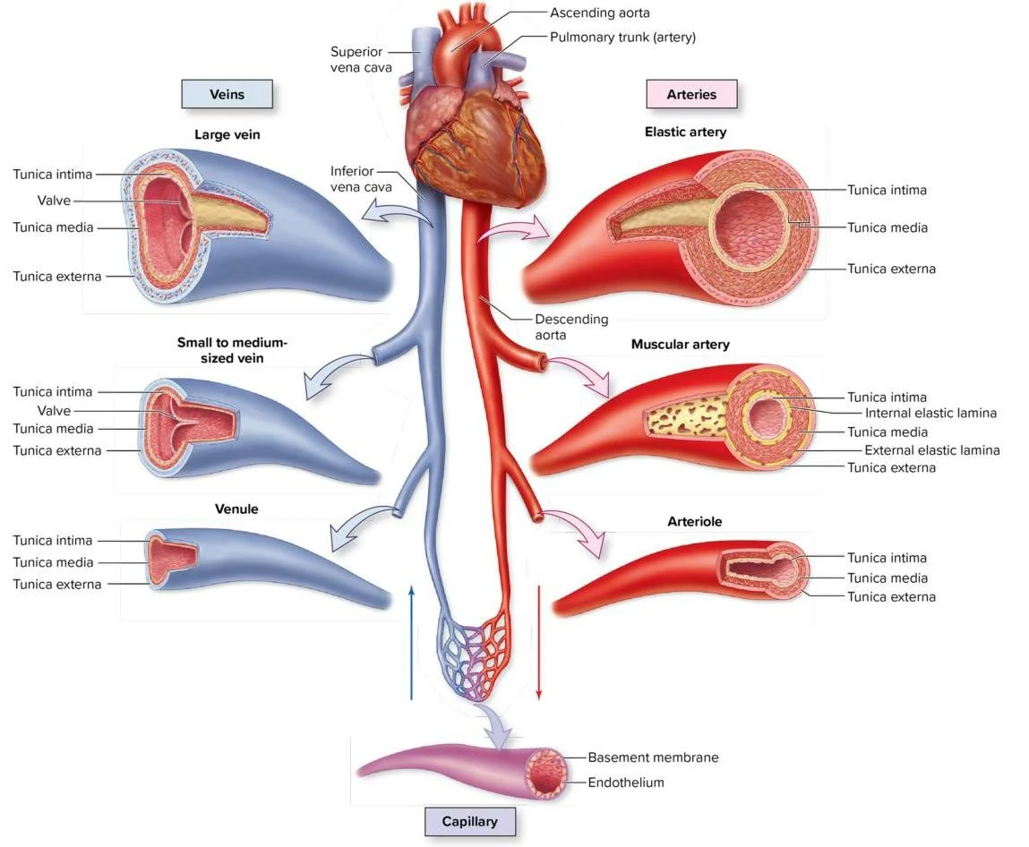
Before diving into specific regional anatomy, it is essential to understand the structural differences between the two main types of vessels. Arteries are "resistance vessels" that carry blood away from the heart under high pressure. Conversely, veins are "capacitance vessels" that return blood to the heart and serve as reservoirs for the majority of the body's blood volume.
The Three Tunics
Both arteries and veins are composed of three distinct layers, known as tunics:
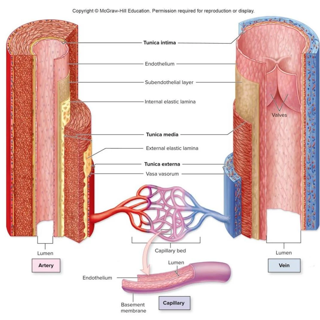
Tunica interna (intima): The innermost layer, consisting of a slick, simple squamous endothelium that acts as a selectively permeable barrier.
Tunica media: The middle layer, composed of smooth muscle, collagen, and elastin. This layer is responsible for vasoconstriction and vasodilation.
Tunica externa (adventitia): The outermost layer of loose connective tissue that anchors the vessel to surrounding structures.
Note on Histology: While arteries have a much thicker tunica media to withstand high pressure, capillaries—the sites of nutrient exchange—consist of only the tunica intima.
Comparison of Vessel Characteristics
Arteries and veins differ significantly in their physical properties. Arteries generally have narrower lumens and thicker walls than their companion veins. Because veins operate under much lower pressure, they often contain valves to prevent the backflow of blood, a feature not found in systemic arteries.
Diagram illustrating the cross-sections of an artery, vein, and capillary, detailing their respective layers and structures, including the tunica intima, tunica media, tunica externa, endothelium, basement membrane, and valves in veins.Diagram illustrating the structure of the human circulatory system, showing the heart, arteries, veins, arterioles, venules, and capillaries, along with cross-sections detailing their respective layers.
Arteries of the Head, Neck, and Face
The blood supply to the head and neck is highly specialized to ensure the brain receives a constant, protected flow of oxygenated blood.
The Carotid System
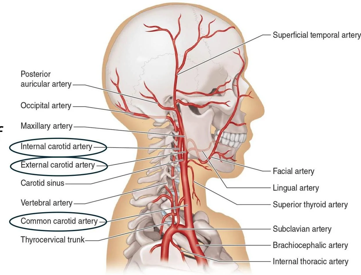
The common carotid arteries are the primary vessels supplying this region. The left common carotid arises directly from the aorta, while the right arises from the brachiocephalic trunk. As these vessels ascend, they bifurcate into two distinct branches:
External Carotid Artery: This branch supplies the external structures, including the face, neck muscles, tongue, teeth, sinuses, and ears.
Internal Carotid Artery: This branch travels deeper to supply the interior cranium, specifically the brain and the pituitary gland.
An illustration of the arteries in the human head and neck, including the common carotid artery, internal carotid artery, external carotid artery, and their branches.
Posterior Circulation and the Circle of Willis
In addition to the carotid system, the vertebral arteries ascend through the transverse foramina of the vertebrae to supply the spinal cord, vertebrae, and cerebellum. The left and right vertebral arteries merge to form the basilar artery.
These various arterial pathways converge to form an anastomosis known as the Circle of Willis. This circular arrangement acts as a safety mechanism; if one vessel becomes blocked, the Circle of Willis provides collateral channels to maintain blood flow to the brain.
Diagram illustrating the arteries of the brain, including the Circle of Willis, vertebral arteries, basilar artery, and their branches.
Trunk and Abdominal Circulation
The central part of the arterial system is dominated by the aorta, the body's largest conducting artery. The aorta is divided into several sections:
Ascending Aorta: Gives rise to the brachiocephalic trunk, which further branches into the right and left subclavian arteries.
Aortic Arch: The curved portion of the vessel.
Descending Aorta: This section provides parietal branches to the thoracic and abdominal skeletal muscles, as well as visceral branches to the internal organs.
As the descending aorta reaches the lower abdomen, it bifurcates into the common iliac arteries, which then split into the internal and external iliac arteries to supply the pelvis and lower extremities.
Venous Return of the Trunk
The venous system of the trunk is organized to return blood to the heart via two massive vessels:
Superior Vena Cava: Formed from the brachiocephalic veins, it drains blood from the head, neck, upper extremities, and parts of the thorax and abdomen.
Inferior Vena Cava: Formed from the common iliac veins, it drains the lower extremities, pelvis, and much of the abdomen.
Arteries and Veins of the Upper Extremity
The vessels of the upper limb follow a continuous path from the shoulder to the fingertips, changing names as they pass specific anatomical landmarks.
Arterial Pathway
The arterial flow follows this sequence:
Subclavian Artery→ (passes lateral border of 1st rib) →Axillary Artery→ (passes inferior to teres major) →Brachial Artery→ (splits in cubital fossa) →Radial and Ulnar Arteries.
The radial artery supplies the anterior lateral forearm and hand, while the ulnar artery supplies the medial forearm and hand. In the palm, these arteries merge to form the superficial and deep palmar arches, which then give rise to the digital arteries that supply the fingers.
An illustration of the arteries of the human arm and hand, including the subclavian, axillary, brachial, radial, and ulnar arteries, as well as the volar arches and digital arteries.
Venous Drainage and Clinical Application
The veins of the upper extremity are categorized into deep and superficial layers.
Deep Veins: These generally parallel the arteries and share their names (e.g., brachial, radial, and ulnar veins).
Superficial Veins: These include the cephalic, basilic, and median veins.
Clinical Tip: Venipuncture
The median cubital vein, which connects the cephalic and basilic veins in the elbow (cubital fossa) area, is a common site for venipuncture (drawing blood) because it is superficial and easily accessible.
Summary of Clinical Importance
Understanding these vessels is vital for several clinical reasons:
Pulse Points: The radial artery is the most common site for measuring heart rate, while the brachial artery is the standard site for blood pressure measurements. The carotid artery also serves as an important pulse point.
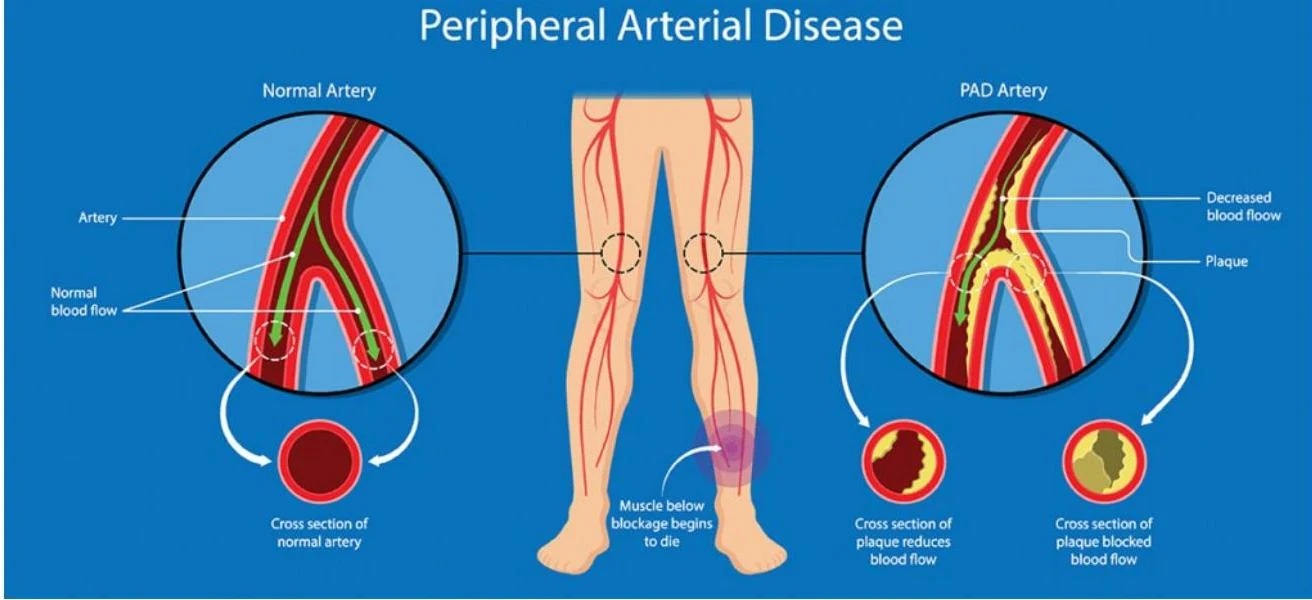
Vascular Disease:Peripheral Artery Disease (PAD) involves the narrowing of arteries in the limbs due to atherosclerosis, leading to pain and numbness. In the venous system, varicose veins occur when valves become incompetent, causing blood to pool and veins to swell.
This illustration compares a normal artery with an artery affected by Peripheral Arterial Disease (PAD), showing plaque buildup and reduced blood flow.
Keep reading for free.
Create an account to finish this reading and make it part of your study routine.