Explores the intricate anatomy of the human eye, detailing its structures such as the cornea, iris, lens, and retina. It explains the physiological processes of vision, including light refraction, accommodation, pupillary reflexes, and phototransduction within the rods.
The human eye is a sophisticated biological instrument designed to capture light, refract it to a precise focal point, and convert that electromagnetic energy into electrical signals that the brain can interpret. This process involves a seamless integration of anatomical structures, mechanical adjustments, and complex biochemical cascades. Understanding vision requires looking at the eye not just as a camera, but as a dynamic sensory organ that constantly adjusts to its environment.
Anatomy of the Eye
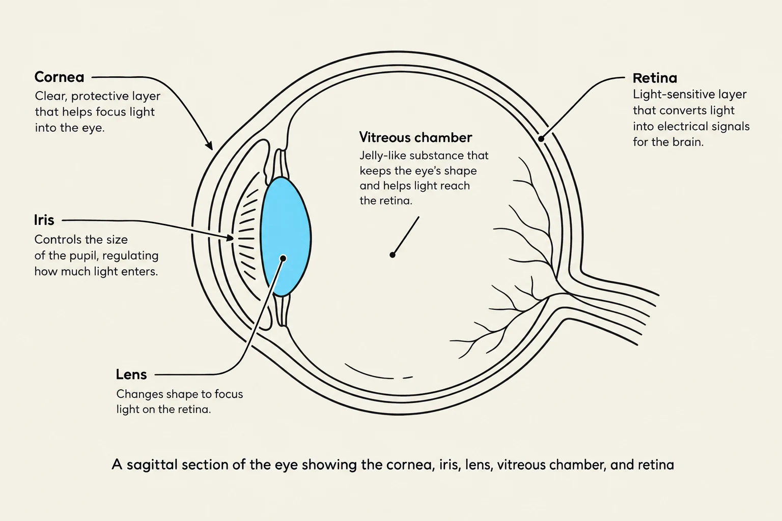
The eye is composed of several distinct layers and chambers that work together to manage light entry and focus. The outermost layer provides structural integrity and protection. The sclera is a tough, white connective tissue that forms the bulk of the eyeball. At the front of the eye, the sclera transitions into the cornea, a transparent structure that plays a primary role in the initial refraction (bending) of light as it enters the eye.
Inside the eye, the space is divided into two main segments. The anterior segment contains the aqueous humor, a clear fluid that maintains intraocular pressure and provides nutrients. This fluid is regulated by the Canal of Schlemm, which helps drain the humor to maintain balance. The posterior segment is occupied by the vitreous chamber, which is filled with a gel-like substance that helps maintain the eye's spherical shape.
A sagittal section of the eye showing the cornea, iris, lens, vitreous chamber, and retina.
The light then passes through the pupil, which is the central opening of the iris. The iris acts as a diaphragm, adjusting the size of the pupil to control the amount of light entering the eye. Behind the iris lies the lens, a flexible structure responsible for fine-tuning the focus of light onto the back of the eye. The lens is held in position by zonules, which are inelastic ligaments that connect the lens to the ciliary muscle.
The innermost layer is the retina, the sensory tissue where light is converted into neural signals. Within the retina, the macula serves as a specialized area for central vision, and at its very center is the fovea, the point of highest visual acuity. Blood supply to these delicate tissues is provided by the central retinal artery and vein. Where the axons of the retinal ganglion cells exit the eye, they form the optic disk, often referred to as the "blind spot" because it lacks photoreceptors.
Accommodation and the Pupillary Reflex
Vision is not a static process; the eye must constantly adapt to changing circumstances. One such adaptation is accommodation, the physiological process by which the eye adjusts the shape of the lens to maintain focus on objects at varying distances. When viewing near objects, the ciliary muscle contracts, which changes the tension on the zonules and allows the lens to become more rounded, increasing its refractive power.
Another vital reflex is the pupillary reflex, which regulates light intensity. When bright light hits the retina, the pupil constricts to protect the retina from overexposure. This reflex is regulated by Cranial nerve III (the oculomotor nerve), which sends signals to the muscles of the iris to induce constriction.
Study Tip: Remember the relationship between the ciliary muscle and the lens. In accommodation, the ciliary muscle contracts to make the lens rounder for near vision. This is the opposite of how many other muscles work in the body!
Neural Pathways for Vision
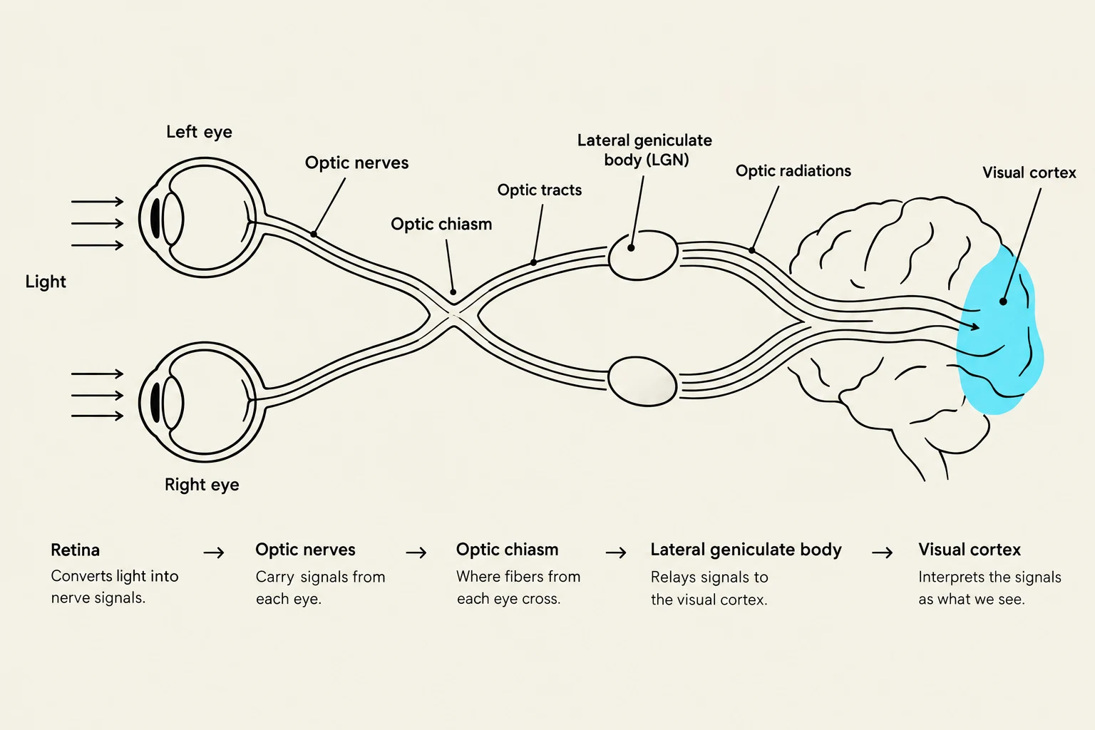
Once the retina captures light, the information must be transmitted to the brain. The electrical impulses travel from the retina through the optic nerve. These nerves meet at the optic chiasm, where some nerve fibers cross to the opposite side of the brain, allowing for binocular vision.
From the chiasm, the fibers continue as the optic tract toward the thalamus. Here, they synapse in the lateral geniculate body, which acts as a relay station. While most signals proceed to the visual cortex located in the occipital lobe for high-level processing, collateral pathways also lead to the midbrain to facilitate reflexes like the pupillary reflex.
Diagram illustrating the pathway of visual information from the eyes to the visual cortex of the brain, including the optic nerves, optic chiasm, optic tracts, and lateral geniculate body.
Phototransduction in Rods
The most complex part of vision is phototransduction, the process of converting light into a change in membrane potential. While the eye has various receptors, the rods are specialized for low-light (scotopic) vision.
In total darkness, the photoreceptor cells are actually in a relatively "active" state regarding neurotransmitter release. The pigment rhodopsin is inactive, which allows levels of cyclic GMP (cGMP) to remain high within the cell. High cGMP levels keep cyclic nucleotide-gated (CNG) and $K^+$ channels open. This constant influx of positive ions maintains a relatively depolarized membrane potential of approximately −40mV, which allows for the tonic (continuous) release of neurotransmitters onto bipolar neurons.
When light strikes the rod, the following cascade occurs:
Activation: Light "bleaches" the rhodopsin, changing its shape and activating it.
G-Protein Activation: The activated rhodopsin triggers a G-protein called transducin.
cGMP Reduction: Transducin initiates a cascade that decreases the concentration of cGMP.
Channel Closure: As cGMP levels drop, the CNG channels close.
Hyperpolarization: With the positive ions no longer entering the cell, the membrane potential drops (becomes more negative), hyperpolarizing to approximately −70mV.
Signal Transmission: This hyperpolarization causes a decrease in the release of neurotransmitters onto the bipolar neurons, which the brain interprets as a light signal.
State
Rhodopsin
cGMP Levels
CNG Channels
Membrane Potential
Neurotransmitter Release
Darkness
Inactive
High
Open
≈−40mV (Depolarized)
High (Tonic)
Light
Activated
Low
Closed
≈−70mV (Hyperpolarized)
Decreased
Keep reading for free.
Create an account to finish this reading and make it part of your study routine.