Detailed explanation of the anatomical structures and physiological functions of the human eye, covering vision, and the human ear, covering hearing and balance. Includes details on photoreceptors, visual pathways, auditory pathways, and equilibrium mechanisms.
The human body relies on specialized sensory organs to interpret the external world. Among the most complex of these are the eyes and the ears. While the eyes function as photoreceptors to translate light into visual images, the ears serve a dual purpose: acting as mechanoreceptors for audition (hearing) and as the primary organs for maintaining equilibrium (balance). Understanding the intricate anatomy of these organs is essential for grasping how neural impulses are generated and processed by the brain.
The Eye: Anatomy and Vision
The eyeball is a sophisticated organ composed of three distinct layers, known as tunics, which protect the internal structures and facilitate the refraction of light.
The Eye Tunics
The external wall of the eye is organized into three layers:
Fibrous Tunic (External): This is the outermost layer. It includes the sclera, a white, opaque coat of dense connective tissue that provides shape and protection, and the cornea, a transparent, avascular layer at the anterior pole that allows light to enter.
Vascular Tunic (Middle/Uvea): This layer contains the choroid, which provides a vast network of capillaries to supply nutrients and oxygen to the retina. It also includes the ciliary body, which contains muscles that alter the shape of the lens, and the iris, the colored part of the eye that regulates light entry through the pupil.
Sensory Tunic (Internal): The retina is the innermost layer. It consists of an outer pigmented layer and an inner neural layer that houses specialized photoreceptors.
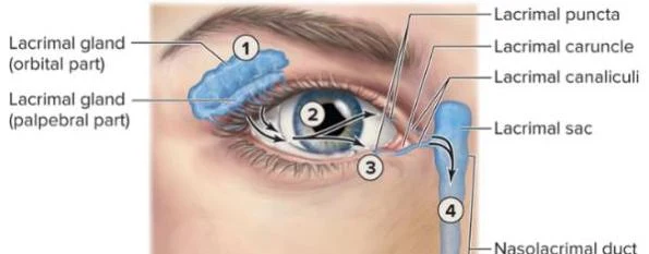
Diagram illustrating the anatomy of the human eye and the lacrimal system, showing the lacrimal gland, lacrimal puncta, lacrimal caruncle, lacrimal canaliculi, lacrimal sac, and nasolacrimal duct.Anatomical illustration of the human eye and its surrounding structures, including muscles, nerves, and eyelid components.
Photoreceptors and the Retina
The retina converts light energy into nerve impulses using two types of photoreceptor cells:
Rod cells: Responsible for vision in dim light and perceiving shades of gray.
Cone cells: Responsible for color vision and high acuity in bright light.
The area of highest visual resolution is the fovea centralis, located at the center of the macula lutea, which contains primarily cones.
The Visual Pathway and Interior
The interior of the eye is divided by the lens into two cavities:
Anterior Cavity: Filled with aqueous humor, which maintains intraocular pressure to prevent the eye from collapsing.
Posterior Cavity: Filled with vitreous humor, a jelly-like substance that presses the retina against the choroid.
Visual information follows a specific pathway: light is captured by the retina, converted to impulses by the optic nerve (CN II), crosses at the optic chiasm, travels through the lateral geniculate body (LGB) of the thalamus, and finally reaches the visual cortex in the occipital lobe.
Exam Tip: Remember that the optic disc is the location where the optic nerve exits the eye. Because it lacks photoreceptors, it is known as the "blind spot".
Extrinsic Eye Muscles and Age-Related Issues
To coordinate movement between the two eyes, six extrinsic eye muscles (such as the superior rectus and lateral rectus) work in concert. As humans age, several ocular issues may arise:
Presbyopia: The loss of lens elasticity, making it difficult to focus on close objects.
Glaucoma: A condition caused by increased intraocular pressure (often due to aqueous humor buildup), which can lead to irreversible retinal damage and blindness.
The Ear: Hearing and Equilibrium
The ear is divided into three functional regions: the external, middle, and internal ear.
External and Middle Ear
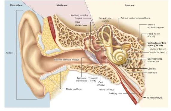
The external ear consists of the auricle (pinna), which collects sound waves, and the external acoustic canal, which directs them toward the tympanic membrane (eardrum).
The middle ear is an air-filled cavity containing the three smallest bones in the body, known as the ear ossicles: the malleus, incus, and stapes. These bones transmit vibrations from the tympanic membrane to the oval window. To prevent damage from loud sounds, the stapedius and tensor tympani muscles can dampen these vibrations. Additionally, the pharyngotympanic (Eustachian) tube connects the middle ear to the nasopharynx to equalize air pressure.
Internal Ear: Cochlea and Equilibrium
The internal ear contains the bony and membranous labyrinths and is responsible for both hearing and balance.
Hearing (The Cochlea)
The cochlea is a snail-shaped structure dedicated to hearing. Within the cochlear duct (scala media) lies the organ of Corti, which contains hair cells. When sound vibrations move the fluid in the cochlea, these hair cells bend, triggering neural impulses.
Equilibrium (The Vestibular Apparatus)
The vestibular apparatus manages our sense of balance through two mechanisms:
Static Equilibrium: Managed by the vestibule (containing the utricle and saccule), which monitors the head's position relative to gravity and detects linear acceleration.
Dynamic Equilibrium: Managed by the semicircular canals, which monitor rotational or spinning movements (angular acceleration).
Diagram of the human ear, illustrating the external, middle, and inner ear structures, including the auricle, auditory ossicles, semicircular canals, cochlea, and auditory tube.
The Auditory Pathway
Auditory signals are transmitted via the vestibulocochlear nerve (CN VIII). The signal travels from the cochlear nuclei in the medulla, through the pons and midbrain, to the medial geniculate body of the thalamus, and finally to the primary auditory area in the temporal lobe of the cerebral cortex.
Clinical Note: Age-related hearing loss, known as presbycusis, often results from the loss of hair cells in the organ of Corti or the degeneration of neural pathways.
Keep reading for free.
Create an account to finish this reading and make it part of your study routine.