Explore the heart's structure, including its chambers, valves, and walls, and understand its role in pumping blood through the pulmonary and systemic circuits. Learn about the cardiac cycle and its regulation.
The heart is the central engine of the cardiovascular system, acting as a nonstop pump that averages about 75 beats per minute. It is responsible for generating the blood pressure necessary to circulate blood through two distinct circuits: the pulmonary circuit, which directs blood to the lungs for gas exchange, and the systemic circuit, which delivers oxygenated blood to the rest of the body.
Location and Protective Structures
The heart is the largest organ of the mediastinum, located between the lungs and slightly to the left of the body's midline. It is roughly the size of a fist and weighs less than one pound. Anatomically, the base of the heart is its superior aspect, while the apex points inferiorly toward the left.
An illustration of the human heart and lungs, showing major anatomical structures like the aorta, pulmonary trunk, superior vena cava, and diaphragm.
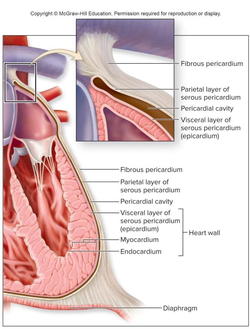
To protect this vital organ, it is enclosed in a tough, double-walled sac called the pericardium. The pericardium consists of several distinct layers:
Fibrous pericardium: The outermost, strong layer of dense connective tissue that is fused to the diaphragm and major vessels.
Serous pericardium: A thinner, two-layered membrane consisting of:
Parietal pericardium: The outer layer of the serous membrane.
Visceral pericardium: The inner layer that sits directly on the heart surface.
Pericardial cavity: The fluid-filled space located between the parietal and visceral layers, which reduces friction during heart contractions.
Diagram illustrating the layers of the heart wall, including the fibrous pericardium, parietal and visceral layers of the serous pericardium, pericardial cavity, myocardium, endocardium, and diaphragm.
The Heart Wall and Cardiac Muscle
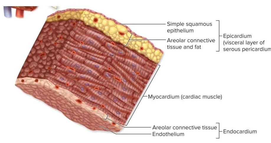
The heart wall itself is composed of three distinct layers:
Epicardium: The superficial layer, which is also the visceral layer of the serous pericardium.
Myocardium: The thick middle layer composed of cardiac muscle tissue. This muscle is striated (similar to skeletal muscle) and arranged in a complex vortex or spiral pattern. This arrangement is crucial for the efficient pumping of blood.
Endocardium: The deep layer that lines the internal chambers of the heart.
Diagram illustrating the layers of the heart wall: epicardium, myocardium, and endocardium.
Histology of Cardiac Muscle
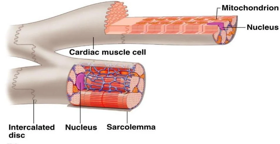
Cardiac muscle cells are characterized by being relatively short, branched, and containing only one or two nuclei. A defining feature of this tissue is the presence of intercalated discs, which are complex junctions that allow cells to join together and form a functional cellular network. These cells are separated by a delicate endomysium that contains necessary blood vessels and nerves.
Diagram illustrating the structure of a cardiac muscle cell, highlighting its nucleus, mitochondria, sarcolemma, and intercalated discs.
Heart Chambers and Blood Flow
The heart is divided into four hollow chambers: two superior atria and two inferior ventricles.
The Right Side (Pulmonary Circuit)
The right side of the heart manages deoxygenated blood:
Right Atrium: Receives venous blood from the systemic circuit via the superior vena cava, inferior vena cava, and coronary sinus. It features a "C" shaped ridge called the crista terminalis.
Right Ventricle: Receives blood from the right atrium and pumps it into the pulmonary trunk toward the lungs. Its internal walls feature irregular ridges known as trabeculae carneae.
The Left Side (Systemic Circuit)
The left side handles oxygen-rich blood:
Left Atrium: Receives oxygenated blood from the lungs via the pulmonary veins.
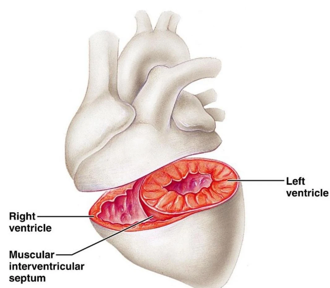
Left Ventricle: Pumps blood through the aortic semilunar valve into the ascending aorta to supply the entire body. Notably, the left ventricle is three times thicker than the right ventricle because it must exert much greater force to overcome the resistance of the systemic circuit.
An illustration of the human heart, highlighting the right ventricle, left ventricle, and the muscular interventricular septum.
Heart Valves and the Fibrous Skeleton
To ensure that blood flows in only one direction, the heart utilizes four valves:
Atrioventricular (AV) Valves
These valves are located between the atria and the ventricles and close when the ventricles contract to prevent backflow into the atria:
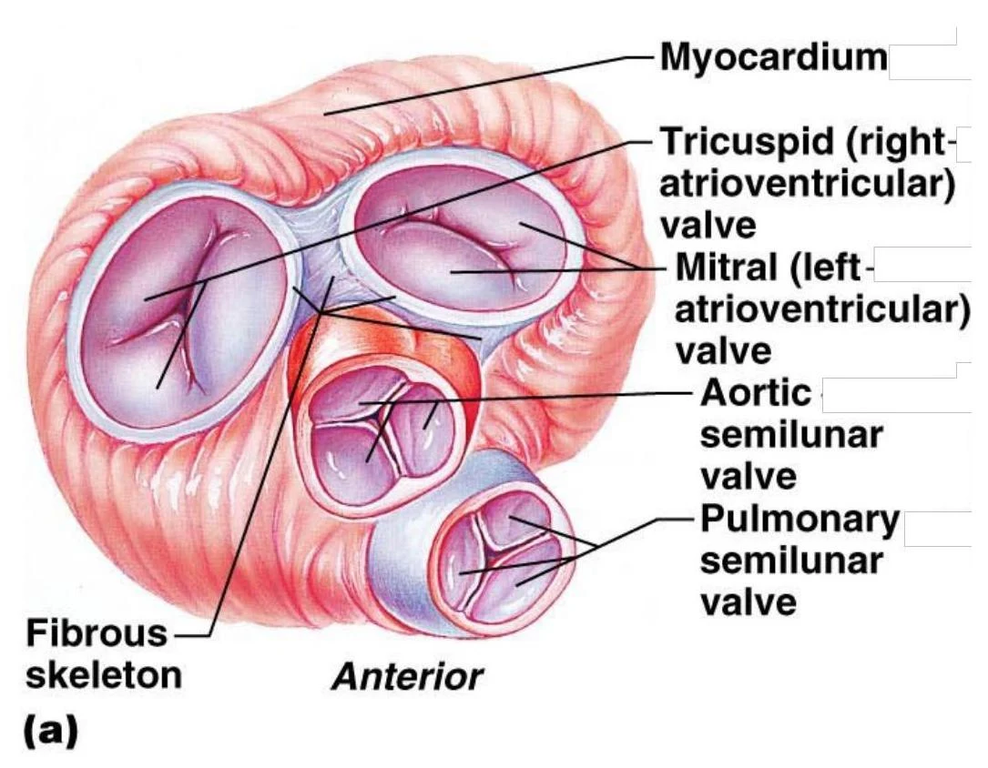
Tricuspid Valve: Located between the right atrium and right ventricle; it has three cusps.
Mitral (Bicuspid) Valve: Located between the left atrium and left ventricle; it has two cusps.
These AV valves are supported by chordae tendineae—tendon-like structures attached to the free edges of the cusps—which prevent the valves from prolapsing into the atria during contraction.
Semilunar Valves
These valves are located at the exits of the ventricles and close when the ventricles relax:
Pulmonary Semilunar Valve: Located between the right ventricle and the pulmonary trunk.
Aortic Semilunar Valve: Located between the left ventricle and the ascending aorta.
Unlike AV valves, semilunar valves do not have chordae tendineae. All four valves are anchored by the fibrous skeleton, a structure of dense connective tissue that provides attachment points for the valves, prevents over-dilation of the openings, and acts as an electrical insulator to block the direct spread of impulses between chambers.
An illustration of the human heart valves, including the tricuspid, mitral, aortic semilunar, and pulmonary semilunar valves, along with the myocardium and fibrous skeleton.
Heart Sounds and the Cardiac Cycle
The rhythmic "lub-dup" sound heard during a heartbeat is produced by the closing of the valves:
"Lub": The first sound, caused by the closing of the AV valves.
"Dup": The second sound, caused by the closing of the semilunar valves.
The cardiac cycle refers to one complete sequence of contraction (systole) and relaxation (diastole).
Exam Tip: Remember that the "lub" sound is associated with the AV valves (tricuspid/mitral) and the "dup" sound is associated with the semilunar valves (aortic/pulmonary).
Regulation and Blood Supply
While the heart has an intrinsic rhythm, the autonomic nervous system modulates its activity:
Sympathetic division: Stimulates the myocardium to increase the force of contraction.
Parasympathetic division: Slows the heart rate via the vagus nerves.
The heart muscle itself requires a dedicated blood supply provided by the coronary arteries, which arise from the aorta. Venous drainage of the heart is primarily handled by the coronary sinus, which returns about 80% of the blood to the right atrium.
Clinical Note: As humans age, arteries can stiffen and the heart muscle may thicken, which can reduce chamber volume and overall cardiac output. Furthermore, coronary artery disease—specifically atherosclerosis (fatty deposits)—is a leading cause of myocardial infarction (heart attack).
Keep reading for free.
Create an account to finish this reading and make it part of your study routine.