


The heart is a complex muscular organ composed of several specialized chambers, valves, and vessels that facilitate systemic and pulmonary circulation. To understand how the heart functions as a pump, one must master its anatomical components, ranging from the large vessels that transport blood to the microscopic muscular tissue that powers every contraction.
The Chambers and Septa
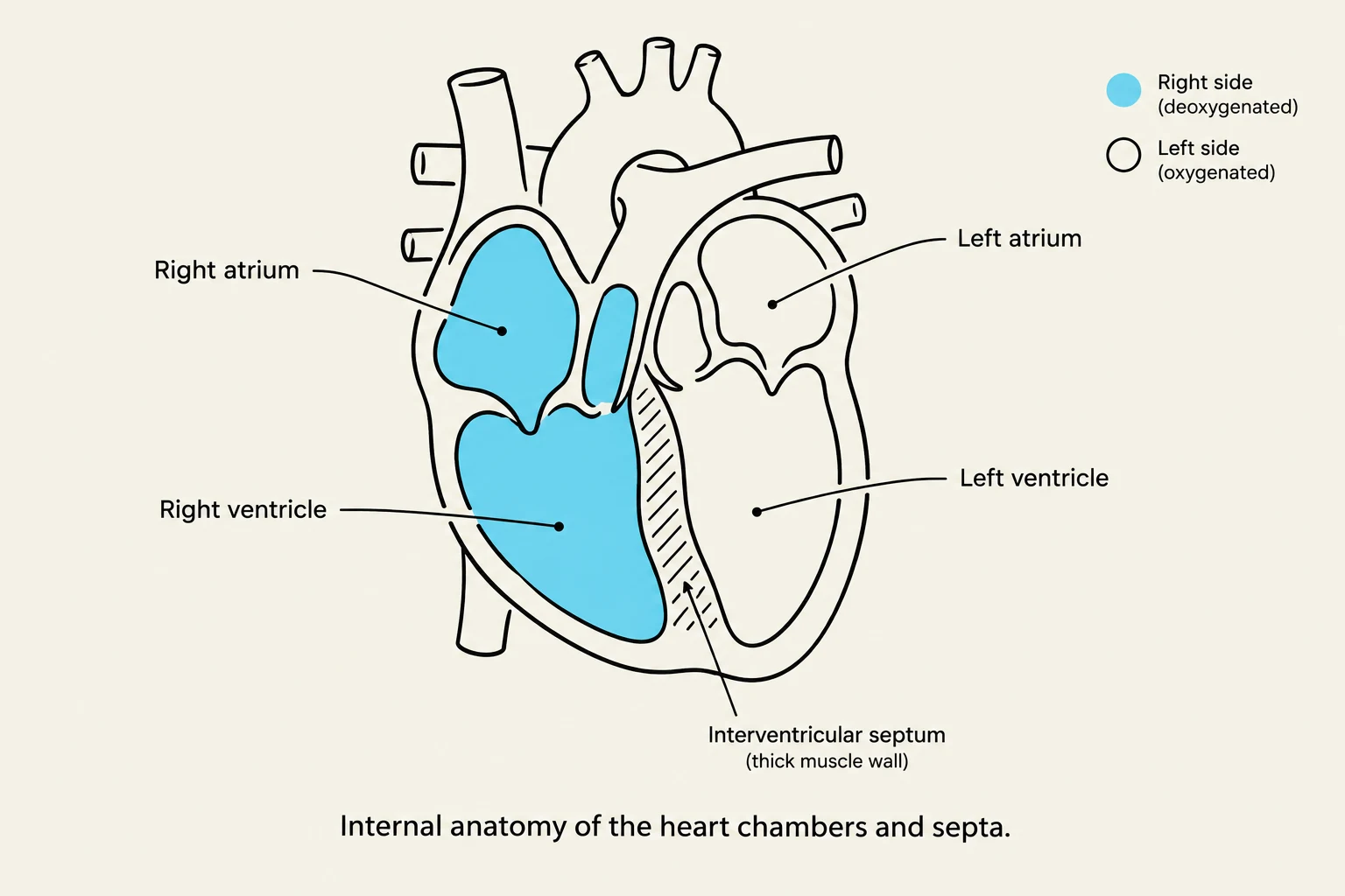
To ensure that oxygenated and deoxygenated blood do not mix, the heart utilizes a thick muscular wall known as the interventricular septum. This separation is vital for maintaining the efficiency of gas exchange in the lungs and nutrient delivery to the tissues. Within the atria, you may also observe pectinate muscles, which are muscular ridges that aid in contraction.
Exam Tip: When identifying structures on a diagram, remember that the left side of the heart (the left atrium and left ventricle) handles oxygenated blood, while the right side handles deoxygenated blood.
The Pathway of Blood Flow
Understanding the heart requires following the continuous loop of blood through various vessels and valves.
The Pulmonary Circuit (Deoxygenated Blood)
Deoxygenated blood enters the heart through the superior vena cava and inferior vena cava into the right atrium. From there, it passes through the tricuspid valve into the right ventricle. The right ventricle then pumps this blood through the pulmonary semilunar valve into the pulmonary trunk. The trunk branches into the right pulmonary artery and left pulmonary artery, which carry the blood to the lungs to receive oxygen.
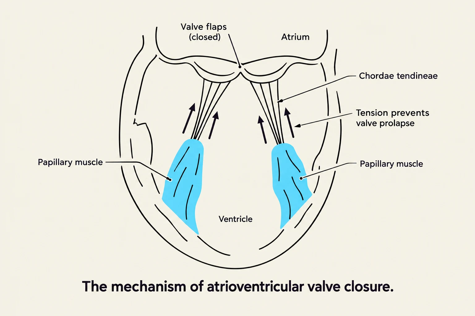
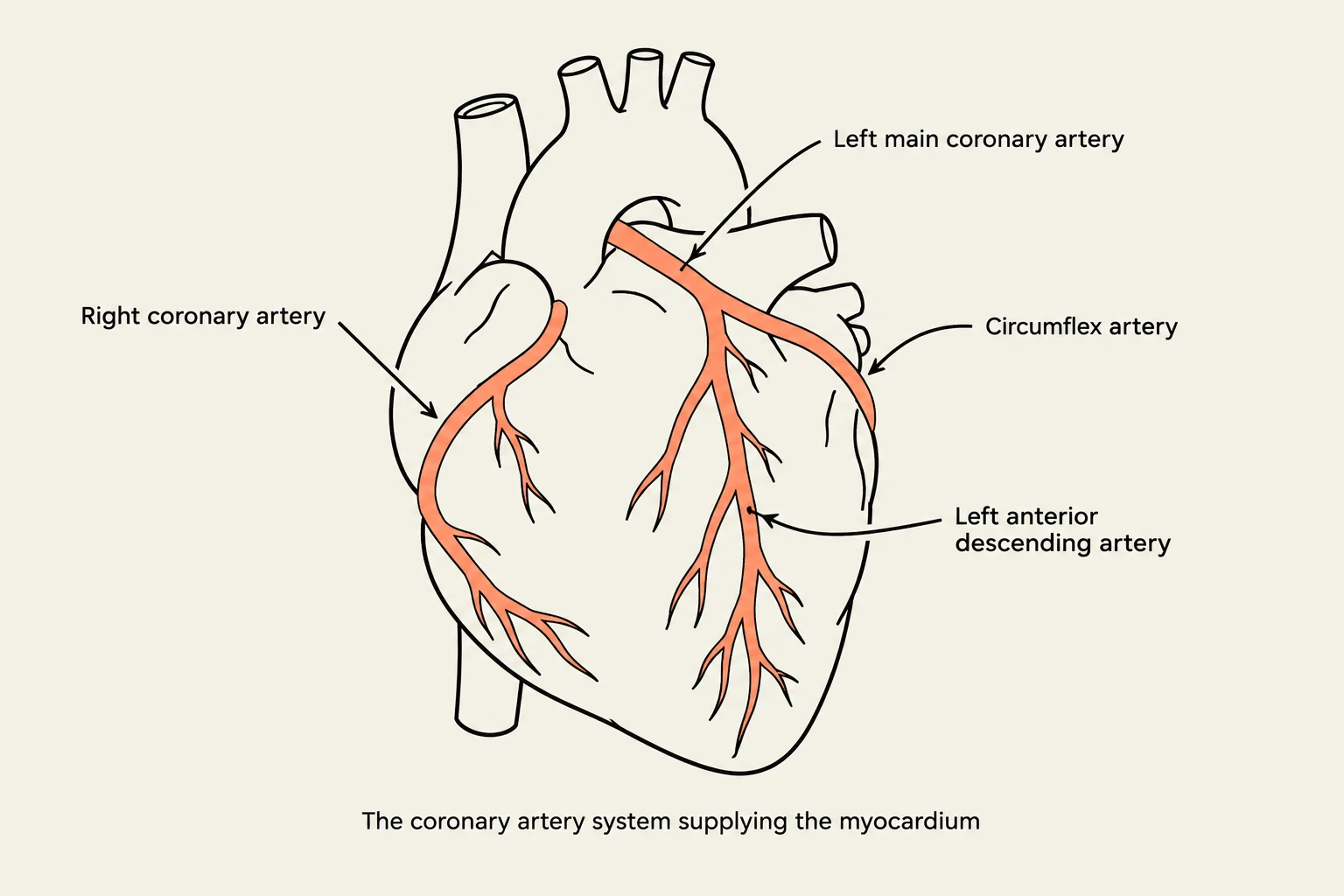
Keep reading for free.
Create an account to finish this reading and make it part of your study routine.
Continue reading freeAlready have an account? Log in
Updated