Anatomy and Physiology of the Female Reproductive System
A comprehensive overview of the female reproductive system, covering its anatomy, the processes of oogenesis and folliculogenesis, hormonal regulation, the menstrual cycle, fertilization, gestation, and common pathophysiological conditions like endometriosis and gestational diabetes.
The female reproductive system is a complex, highly regulated network of organs and hormones designed to facilitate reproduction, support pregnancy, and manage the menstrual cycle. Understanding this system requires a deep dive into its anatomical structures, the intricate cellular processes of egg development, and the hormonal feedback loops that govern everything from ovulation to childbirth.
Anatomy of the Female Reproductive System
The female reproductive system is divided into external and internal structures, each serving specific roles in sexual response, protection, and reproduction.
External Genitalia (The Vulva)
The external structures, collectively known as the vulva, include the mons pubis (a fatty tissue pad), the labia majora (outer skin folds for protection), and the labia minora (inner folds). Other key components include the clitoris, which is highly innervated for sensory response, the urethral opening, and the vaginal orifice.
Internal Reproductive Tract
The internal organs are responsible for the actual processes of fertilization and gestation:
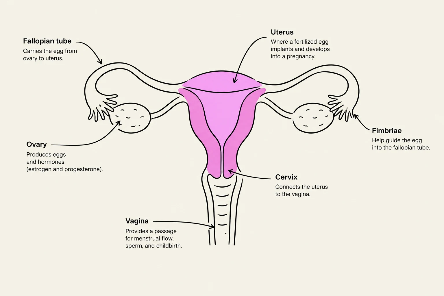
Vagina: A canal with an acidic pH (approximately 3.8 to 4.5) that helps prevent infection.
Cervix: The "neck" of the uterus that connects to the vagina; its mucus consistency changes during the cycle to assist sperm movement.
Uterus: A muscular organ with three distinct layers: the endometrium (the innermost layer that thickens for implantation and sheds during menstruation), the myometrium (the thick smooth muscle layer that contracts during labor), and the fundus (the top portion).
Fallopian Tubes (Uterine Tubes): These tubes connect the ovaries to the uterus. They consist of the infundibulum (funnel-shaped end with finger-like fimbriae), the ampulla (the widest section and the most common site for fertilization), and the isthmus (the narrowest part connecting to the uterus).
Ovaries: Paired, almond-shaped organs that house the follicles. They consist of an outer cortex (containing follicles) and an inner medulla (containing blood vessels and nerves).
Diagram of the female reproductive system, labeling the fallopian tube, ovary, uterus, cervix, vagina, and fimbriae.
Oogenesis and Folliculogenesis
Reproduction relies on two simultaneous processes: oogenesis (the development of the egg/oocyte) and folliculogenesis (the maturation of the ovarian follicle surrounding the egg).
Fetal Development and Atresia
Before birth, primordial germ cells migrate to the ovaries to become oogonia, which replicate via mitosis. These oogonia transform into primary oocytes and enter Meiosis I, where they arrest at prophase I until puberty.
A critical concept in women's health is atresia—the natural degeneration and absorption of ovarian follicles. While a female is born with approximately 1 million primary oocytes, many undergo atresia before birth, leaving only about 300,000 by the time puberty is reached.
Puberty and Hormonal Regulation
At puberty, the hypothalamus releases GnRH, stimulating the anterior pituitary to release Follicle-Stimulating Hormone (FSH) and Luteinizing Hormone (LH).
FSH: Stimulates the growth and maturation of ovarian follicles.
LH: Triggers the final maturation of the dominant follicle and induces ovulation.
As follicles mature from primary to secondary and finally to tertiary (Graafian) follicles, they develop a fluid-filled antrum and specialized layers like the zona pellucida (which protects the oocyte and prevents polyspermy) and theca cells (which produce androgens converted to estradiol).
Exam Tip: Remember the feedback loops. Developing follicles secrete estrogen (estradiol), which initially exerts negative feedback on LH and FSH to prevent too many follicles from maturing. However, a massive surge in estrogen eventually triggers positive feedback, leading to the LH surge that causes ovulation.
The Menstrual Cycle and Conception
The menstrual cycle (averaging 28 days) coordinates the preparation of the uterus for potential pregnancy:
Menstrual Phase (Days 1-5): Low estrogen/progesterone levels cause the endometrium to shed.
Follicular Phase (Days 1-13): FSH stimulates follicle growth, increasing estrogen and thickening the endometrium.
Ovulation (Day 14): An LH surge causes the Graafian follicle to release a secondary oocyte.
Luteal Phase (Days 15-28): The ruptured follicle becomes the corpus luteum, secreting progesterone to maintain the uterine lining.
Fertilization and Implantation
If sperm is present, fertilization typically occurs in the ampulla of the fallopian tube. The sperm undergoes capacitation, penetrates the zona pellucida, and fuses with the egg's membrane. This triggers the egg to complete Meiosis II and form a zygote.
The zygote undergoes cleavage to become a blastocyst as it travels to the uterus. Implantation occurs in the endometrium around days 5-7. Once implanted, the trophoblast cells produce human chorionic gonadotropin (hCG).
Clinical Correlation: Pregnancy tests work by detecting hCG in urine using monoclonal antibodies. A visible color change or line indicates the presence of this hormone, signaling a successful implantation.
Gestation, Parturition, and Pathophysiology
Gestation and Parturition
Gestation is the ~40-week period of fetal development. It is divided into three trimesters, moving from organogenesis to rapid fetal growth.
Parturition (childbirth) is driven by a positive feedback loop: as the baby descends, cervical pressure triggers oxytocin release, which causes stronger uterine contractions, further stretching the cervix. It is vital to distinguish between Braxton-Hicks contractions (irregular "practice" contractions) and true labor contractions (strong, rhythmic, and progressively intensifying).
Pathophysiological Conditions
Two significant conditions discussed in women's health are endometriosis and gestational diabetes:
Endometriosis: A chronic condition where endometrial-like tissue grows ectopically (outside the uterus), such as on the ovaries or pelvic cavity. Potential causes include retrograde menstruation, immune dysfunction, and genetics. Symptoms include chronic pelvic pain, dysmenorrhea (painful periods), and infertility due to adhesions or scar tissue.
Gestational Diabetes (GDM): This occurs when the body cannot produce enough insulin to overcome the insulin resistance caused by pregnancy hormones, such as human placental lactogen (hPL), estrogen, and cortisol.
Risks to Mother: Increased risk of Type 2 diabetes later and preeclampsia.
Risks to Baby: Macrosomia (large birth weight), preterm birth, and neonatal hypoglycemia.
Keep reading for free.
Create an account to finish this reading and make it part of your study routine.