Anatomy and Physiology of the Cardiovascular System
An in-depth explanation of the cardiovascular system, detailing the structure and function of the heart, blood vessels, and the principles of hemodynamics. Covers cardiovascular diseases and their underlying mechanisms.
The cardiovascular system is a sophisticated transport network essential for maintaining homeostasis. It is composed of two primary components: the heart, which acts as a dual-action pump, and the peripheral blood vessels, which serve as the distribution and collection conduits. Understanding how these components interact—from the microscopic contraction of a single muscle cell to the systemic regulation of blood pressure—is fundamental to the study of human physiology and pathology.
The Anatomy of the Heart
The heart is not merely a muscle; it is a complex organ protected by specialized structures and organized into distinct layers to facilitate efficient pumping.
Protective Layers and the Pericardium
The heart is enclosed within the pericardium, a double-walled sac that also encompasses the roots of the great vessels. This sac consists of a fibrous layer and a serous layer. Between these layers lies the pericardial fluid, a straw-colored liquid that reduces friction during the continuous movement of the heart.
The heart wall itself is composed of three distinct tissue layers:
Epicardium: The outermost serous membrane that serves as a protective layer.
Myocardium: The thick middle layer responsible for contraction.
Endocardium: The innermost layer, consisting of smooth muscle and elastic fibers, which lines the heart chambers and is bathed in blood.
The Microscopic Engine: Cardiomyocytes
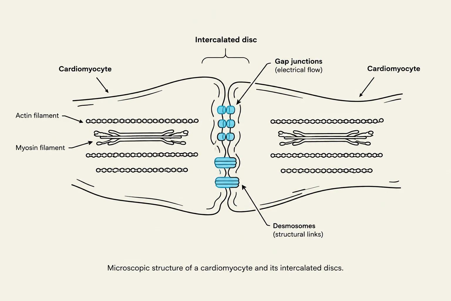
Microscopic structure of a cardiomyocyte and its intercalated discs.
The functional unit of the myocardium is the cardiomyocyte. These specialized cells resemble skeletal muscle but possess electrical properties similar to smooth muscle. To ensure the heart contracts as a single, synchronized unit (a syncytium), cardiomyocytes are joined by intercalated discs. These discs contain:
Gap Junctions: These allow for electronic synchronization, letting electrical impulses pass rapidly between cells.
Desmosomes: These provide structural integrity, preventing the cells from pulling apart during forceful contractions.
Contraction is driven by myofilaments, specifically the thick filament (myosin) and the thin filament (actin). For the myosin heads to attach to actin and trigger a contraction, the cell requires both calcium and ATP. The sarcoplasmic reticulum plays a critical role here by storing calcium ions; when an electrical impulse arrives, calcium is released into the cell to trigger contraction and is subsequently reabsorbed to allow for relaxation.
Blood Flow and Valvular Function
Blood moves through four chambers: the atria (upper chambers that receive blood) and the ventricles (lower chambers that eject blood). To ensure unidirectional flow, the heart utilizes two types of valves.
Atrioventricular (AV) Valves
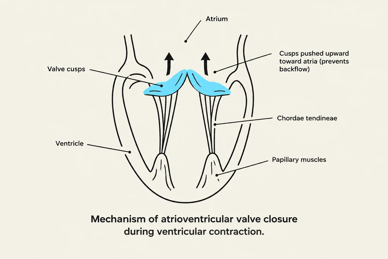
Diagram illustrating the mechanism of atrioventricular valve closure during ventricular contraction, showing valve cusps, chordae tendineae, papillary muscles, atrium, and ventricle.
The AV valves—the tricuspid valve on the right and the mitral (bicuspid) valve on the left—control flow between the atria and ventricles. During ventricular systole (contraction), these valves are forced closed to prevent blood from leaking back into the atria. They are supported by papillary muscles, which are connected to the valve flaps via tendon-like structures called chordae tendineae; these prevent the valves from prolapsing into the atria.
Semilunar Valves
The semilunar valves—the pulmonary valve and the aortic valve—control the exit of blood from the ventricles into the great arteries. These valves close during ventricular diastole (filling) to prevent blood from flowing backward from the arteries into the heart.
Clinical Note: Blood Flow Order
To trace blood through the heart, follow this sequence:
Tricuspid Valve → Pulmonary Valve → Mitral Valve → Aortic Valve.
Hemodynamics: Preload, Afterload, and Ejection Fraction
The efficiency of the heart is measured through several key hemodynamic concepts that describe how much blood is moving and how much resistance the heart faces.
Preload and Starling's Law
Preload refers to the pressure or volume within the ventricles at the end of diastole (the end-diastolic volume). The relationship between volume and force is described by Starling’s Law of the heart, which states that the more the myocardial muscle is stretched by incoming blood, the greater its subsequent force of contraction.
Afterload and Ejection Fraction
Afterload is the resistance that the left ventricle must overcome to eject blood into the systemic circulation. If the heart must constantly pump against excessive resistance (such as in hypertension), it may undergo left ventricular hypertrophy (LVH), where the muscle thickens to compensate.
The ultimate measure of cardiac performance is the Ejection Fraction (EF), which is the percentage of blood ejected from the ventricles with each contraction. A normal EF range is typically between 55-70%.
Peripheral Circulation and Vessel Structure
While the heart provides the pressure, the peripheral blood vessels manage the distribution. Arteries carry blood away from the heart, veins return it, and capillaries facilitate the exchange of gases and nutrients through a single layer of endothelial cells.
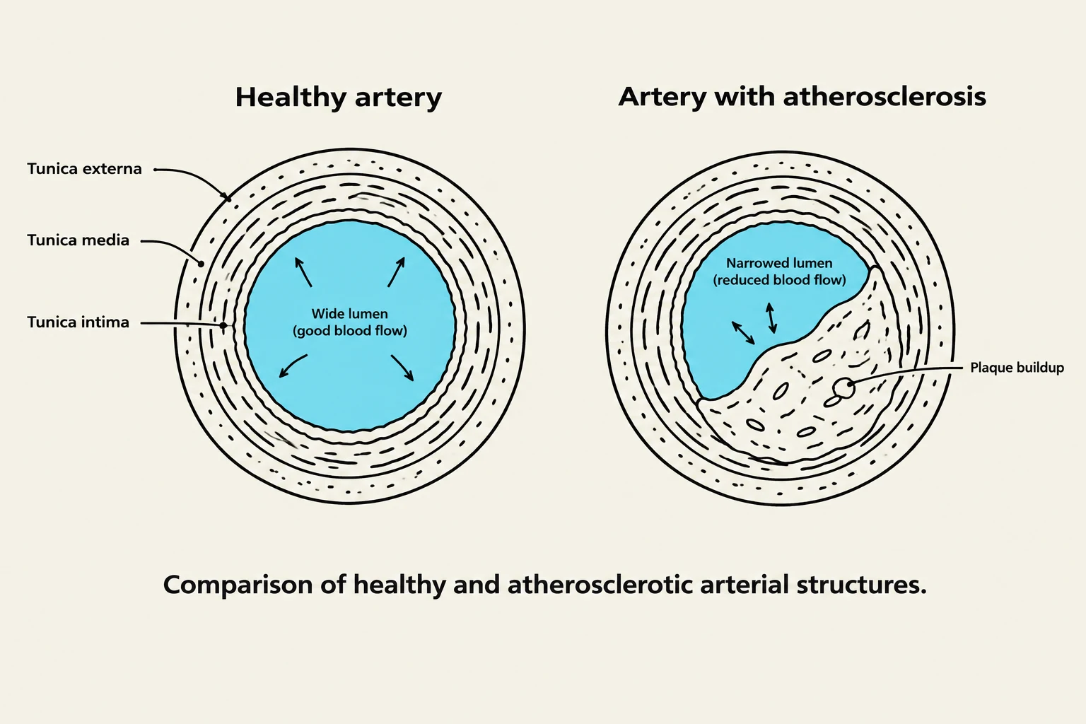
All arteries and veins are composed of three distinct layers:
Tunica Intima: The innermost layer.
Tunica Media: The middle layer, typically containing smooth muscle.
Tunica Externa: The outermost protective layer.
The physics of blood flow is governed by Poiseuille’s Law, which dictates that blood flow is directly proportional to the size of the vessel. To protect against total blockages, the body utilizes anastomoses—communications between two or more vessels—which provide collateral circulation, creating alternative pathways for blood to reach tissues if a primary vessel is obstructed.
Cardiovascular Disease (CVD)
Comparison of healthy and atherosclerotic arterial structures, illustrating plaque buildup and its effect on lumen size and blood flow.
Cardiovascular Disease is a broad term for conditions affecting the heart, the peripheral vessels, or both. Several specific pathologies are common:
Coronary Artery Disease (CAD): A type of CVD involving the arteries that supply blood directly to the heart muscle itself; it is a leading cause of mortality.
Arteriosclerosis: A general disease process characterized by the loss of elasticity in vascular walls due to thickening and hardening.
Atherosclerosis: A specific type of arteriosclerosis where plaque builds up inside the arterial walls, narrowing the lumen.
Exam Tip: Distinguishing Vascular Diseases
Remember that Arteriosclerosis is the broad term for "hardening" of the vessels, while Atherosclerosis refers specifically to the buildup of "plaque" within those vessels.
Summary Table: Hemodynamic Variables
Term
Definition
Clinical Implication
Preload
End-diastolic volume/pressure
Increased stretch increases contraction force (Starling's Law)
Afterload
Resistance to ventricular ejection
High afterload (hypertension) can cause LVH
Ejection Fraction
% of blood ejected per beat
Normal range is 55-70%
Keep reading for free.
Create an account to finish this reading and make it part of your study routine.