This content explains the classification, structure, and function of fibrous, cartilaginous, and synovial joints, along with body movements and examples of specific joints.
Joints, also known as articulations, are the specific sites where two or more bones meet. These connections are essential for the human body because they allow for mobility within the skeleton, enabling everything from delicate finger movements to powerful strides while running.
Classifying Joints
To understand how the body moves, anatomists classify joints using two different systems: structural and functional.
Structural Classifications
Structural classification is based on the material that binds the bones together and whether or not a joint cavity is present:
Fibrous joints: Bones are attached by connective tissue and possess no joint cavity.
Cartilaginous joints: Bones are united by connective tissue and cartilage, also lacking a joint cavity.
Synovial joints: These are characterized by the presence of an obvious joint cavity and are attached by connective tissue and cartilage.
Functional Classifications
Functional classification describes the amount of movement the joint allows:
Synarthroses: Immovable joints.
Amphiarthroses: Slightly movable joints.
Diarthroses: Freely movable joints.
Study Tip: When identifying a joint in a clinical setting, always ask two questions: "What is holding it together?" (Structure) and "How much can it move?" (Function).
Fibrous Joints
Fibrous joints are characterized by bones being joined by fibrous tissue without a joint cavity. Most of these joints are immovable (synarthrotic), though some can be slightly movable (amphiarthrotic).
Common types of fibrous joints include:
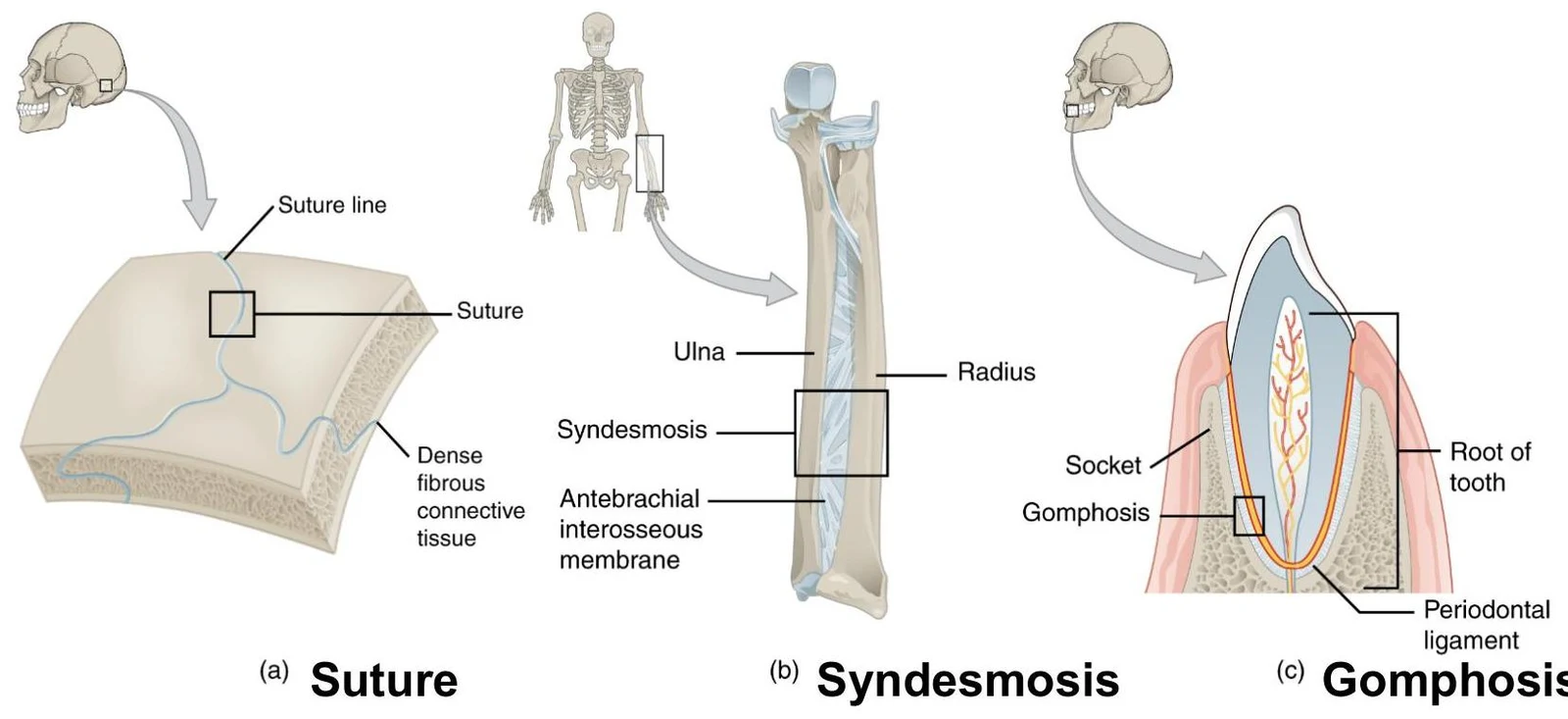
Sutures: Found between the flat bones of the skull.
Gomphoses: The "peg-in-socket" joints of the teeth.
Syndesmosis: A type of joint where bones are connected by a ligament or fibrous membrane. An example of a syndesmosis is the distal articulation between the tibia and fibula, known as the tibiofibular joint. These joints are typically amphiarthrotic.
Diagrams illustrating three types of fibrous joints: (a) Suture, found in the skull; (b) Syndesmosis, between the ulna and radius; and (c) Gomphosis, anchoring a tooth in its socket.
Cartilaginous Joints
In cartilaginous joints, the articulating surfaces are united by cartilage, and no joint cavity is present. Depending on the specific joint, they can be either synarthrotic or amphiarthrotic.
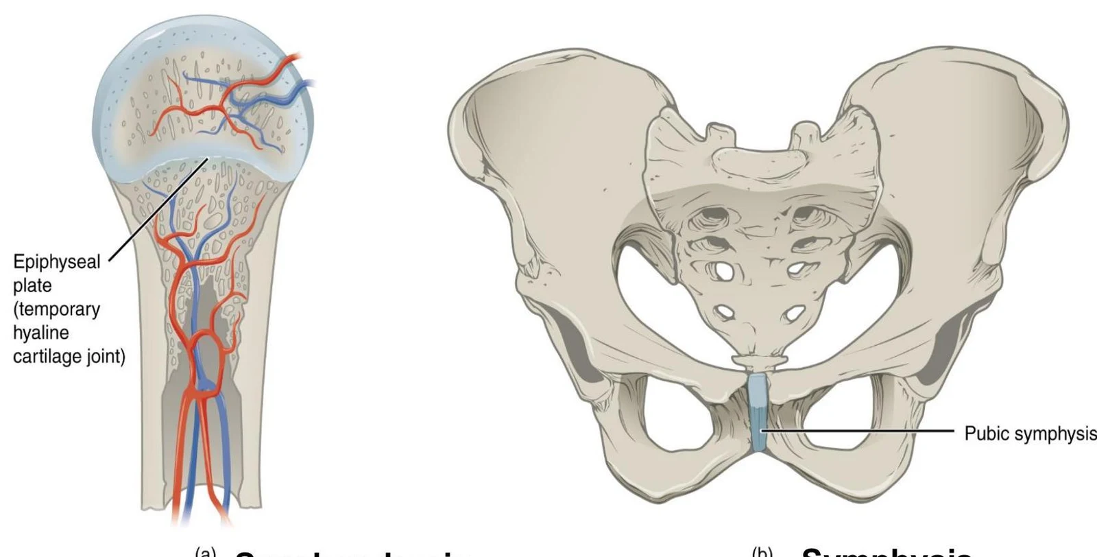
A key type of cartilaginous joint is the symphysis. In a symphysis, the articulating bones are separated by a wedge or pad of fibrocartilage. A common example is the articulation between vertebrae, where a thick pad of fibrocartilage forms the intervertebral disc.
Diagrams illustrating the epiphyseal plate in a long bone and the pubic symphysis of the pelvis.
Synovial Joints
Synovial joints are the most common and mobile joints in the body. They are all classified as diarthrotic (freely movable) and are typically found in the limbs.
Anatomy of a Synovial Joint
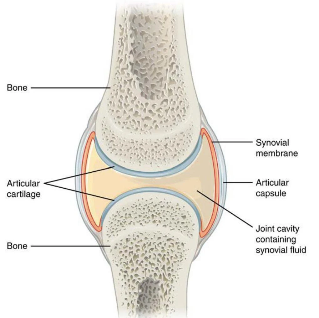
Unlike fibrous or cartilaginous joints, synovial joints feature a fluid-filled joint cavity that separates the articular bones. The key components include:
Articular cartilage: Covers the ends of the bones.
Joint cavity: The space containing the fluid.
Joint capsule: Consisting of an outer articular capsule and an inner synovial membrane.
Synovial fluid: This fluid serves three vital functions: lubrication, nutrient distribution, and shock absorption.
Diagram illustrating the anatomy of a synovial joint, including bones, articular cartilage, synovial membrane, articular capsule, and joint cavity containing synovial fluid.
Types of Synovial Joints and Planes of Motion
Synovial joints are further categorized by the planes and axes of motion they allow:
Joint Type
Description
Motion Axis
Examples
Pivot
A rounded end of one bone fits into a ring of bone or ligament.
A cylindrical projection fits into a trough-shaped surface.
Uniaxial (one plane)
Elbow, knee, ankle, and interphalangeal joints.
Condyloid (Ellipsoid)
An oval articular surface fits into a complementary depression.
Biaxial (two planes)
Radiocarpal (wrist) and metacarpophalangeal (knuckle) joints.
Saddle
Both surfaces have concave and convex areas that interlock.
Biaxial (two planes)
1st carpometacarpal (thumb) and sternoclavicular joints.
Plane (Gliding)
Articular surfaces are flat, allowing short gliding movements.
Multiaxial (more than two planes)
Intercarpal and intertarsal joints.
Ball-and-Socket
A spherical end fits into a cup-like socket.
Multiaxial (more than two planes)
Shoulder and hip joints.
This illustration shows different types of synovial joints in the human skeletal system, including pivot, hinge, saddle, plane, condyloid, and ball-and-socket joints, with examples of their locations in the body.
Body Movements
The movement allowed by a joint is defined by the anatomical planes in which it moves.
Sagittal Plane: Movements like flexion (decreasing the angle of a joint) and extension (increasing the angle) occur here.
Coronal (Frontal) Plane: Movements like abduction (moving away from the midline) and adduction (moving toward the midline) occur in this plane.
Rotation: Turning a bone around its own longitudinal axis (e.g., turning the head side to side).
Circumduction: A circular movement that combines flexion, abduction, extension, and adduction.
Specialized Movements:
Pronation/Supination: Forearm rotation where the radius crosses the ulna (pronation) or they lie parallel (supination).
Dorsiflexion/Plantar Flexion: Moving the foot toward the leg or pointing the toes.
Inversion/Eversion: Turning the sole of the foot toward or away from the midline.
Opposition: The thumb moving to touch the tips of the fingers.
Summary of Selected Joints
To visualize these concepts, consider these specific examples:
The Shoulder (Glenohumeral) Joint: A ball-and-socket joint that provides the widest range of motion in the body.
The Hip Joint: A multiaxial ball-and-socket joint that balances a wide range of motion with high stability.
The Knee Joint: The largest joint in the body, supported by various ligaments (like the ACL and PCL) and the medial/lateral menisci for padding.
The Elbow Joint: A hinge joint that primarily allows for flexion and extension.
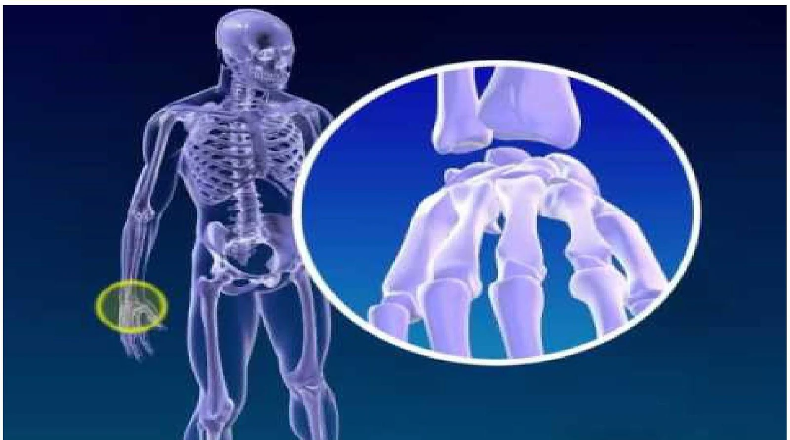
An illustration of the human skeletal structure, with a magnified view of the bones in the wrist and hand.
Note on Development: Joints form during embryonic development alongside the bones. Skull joints typically develop through intramembranous ossification, while limb joints develop through endochondral ossification.
Note on Disorders: Common joint issues include sprains, dislocations, bursitis, and various forms of arthritis.
Keep reading for free.
Create an account to finish this reading and make it part of your study routine.