An overview of the human sensory system, detailing the general and special senses, receptor classifications, and the anatomy of olfaction, gustation, vision, audition, and equilibrium. Includes information on age-related sensory changes.
The human sensory system is a complex network designed to detect changes in the internal and external environments and translate those changes into electrical signals for the brain to interpret. These systems are broadly categorized into general senses and special senses. While general senses are distributed throughout the body to monitor various physiological states, special senses are localized to specific, highly specialized organs.
Receptor Classifications
To understand how we perceive the world, we must first understand the receptors—the specialized structures that detect stimuli. Receptors can be classified in three distinct ways: by their microscopic features, the type of stimulus they detect, and their location in the body.
Microscopic Features
Free nerve endings: These are bare dendrites that detect sensations such as pain, temperature, tickle, itch, and touch.
Encapsulated nerve endings: These consist of dendrites enclosed within a connective tissue capsule, specializing in detecting pressure, vibration, and touch.
Separate cells: Unlike nerve endings, these are distinct receptor cells that synapse with first-order sensory neurons. Examples include the photoreceptors in the retina, the hair cells in the ear, and the gustatory cells in the taste buds.
Stimulus Types
Receptors respond to specific forms of energy:
Photoreceptors: Detect light.
Mechanoreceptors: Detect mechanical pressure or stretching.
Thermoreceptors: Detect changes in temperature.
Osmoreceptors: Detect the osmotic (water) pressure of body fluids.
Chemoreceptors: Detect specific chemicals (e.g., in the nose, mouth, or blood).
Nociceptors: Detect pain resulting from physical or chemical tissue damage.
Location of Stimuli
Exteroceptors: Located near the body surface; they detect external stimuli like smell, vision, taste, touch, and temperature.
Interoceptors: Located within blood vessels and visceral organs; they monitor the internal environment, often felt as pressure or pain.
Proprioceptors: Located in muscles, tendons, and joints; they monitor body position and movement.
Olfaction (Smell)
Olfaction is a special sense that occurs within the olfactory epithelium lining the superior nasal cavity. This tissue contains three major cell types: olfactory receptor cells (which are bipolar neurons), supporting cells that sustain the receptors, and basal cells (stem cells) that undergo mitosis to replace receptors.
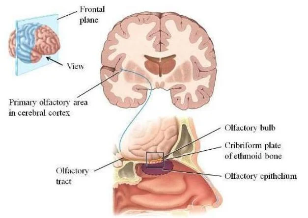
The olfactory receptor cells possess cilia that project from their dendrites; these cilia are the sites where odorant molecules are detected. Once a stimulus is detected, the axons of these neurons gather into bundles that form the olfactory nerve (Cranial Nerve I). These filaments pass through the cribriform plate of the ethmoid bone to reach the olfactory bulbs.
Diagram illustrating the human olfactory system, showing the nasal cavity, olfactory bulb, and the cellular structure of the olfactory epithelium responsible for detecting odor molecules.
From the olfactory bulbs, the axons extend posteriorly as the olfactory tracts (medial and lateral). These tracts synapse in the primary olfactory areas of the temporal and frontal lobes, where smell is perceived. The orbifrontal region is specifically involved in odor identification and discrimination.
This illustration shows the olfactory pathway, including the olfactory bulb, cribriform plate of the ethmoid bone, olfactory epithelium, olfactory tract, and the primary olfactory area in the cerebral cortex.
Gustation (Taste)
Gustation utilizes taste buds to detect chemical stimuli in food. Each taste bud is an oval structure containing three types of epithelial cells: gustatory receptor cells (which feature microvilli called gustatory hairs that project through a taste pore), support cells that form a protective capsule, and basal cells that replace gustatory cells every 7 to 9 days.
Taste Qualities and Papillae
Humans perceive five basic taste qualities: sweet, sour, salty, bitter, and umami (a savory, meaty taste based on protein). These taste buds are housed within different types of tongue papillae:
Fungiform papillae: Mushroom-shaped, located on the tip and sides of the tongue.
Circumvallate (vallate) papillae: Disk-shaped and arranged in a V-shape at the back of the tongue.
Filiform papillae: Small, numerous bumps found throughout the tongue.
Foliate papillae: Located on the lateral margins of the tongue; these typically disappear in early childhood.
The Gustatory Pathway
The innervation of the tongue is divided by region: the anterior 2/3 is served by the facial nerve (CN VII), the posterior 1/3 by the glossopharyngeal nerve (CN IX), and the epiglottis/pharynx by the vagus nerve (CN X). These nerves synapse in the medulla oblongata, after which fibers project to the limbic system, hypothalamus, and thalamus before being integrated in the parietal lobe of the cerebral cortex. Note that the ability to taste relies heavily on the sense of olfaction.
Vision (Sight)
Vision is a complex special sense involving the eyes and several accessory structures designed to protect the ocular surface and ensure it remains moist.
Accessory Structures and the Lacrimal Apparatus
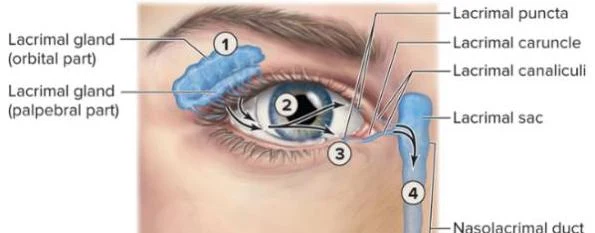
The palpebrae (eyelids) and eyelashes protect the eye from foreign objects, while the eyebrows protect against perspiration and direct sunlight. To maintain moisture, the lacrimal apparatus produces lacrimal fluid (tears) via the lacrimal gland. This fluid contains lysozyme, a bactericidal enzyme that protects the eye. Tears flow over the eyeball, enter the lacrimal puncta, move through the lacrimal canals and sac, and finally drain through the nasolacrimal duct into the nasal cavity.
Diagram illustrating the anatomy of the human eye and the lacrimal system, showing the lacrimal gland, lacrimal puncta, lacrimal caruncle, lacrimal canaliculi, lacrimal sac, and nasolacrimal duct.
Anatomy of the Eyeball
The eyeball is composed of three external tunics:
Fibrous Tunic: The outermost layer, consisting of the transparent cornea (which receives oxygen from lacrimal fluid and aqueous humor) and the white, opaque sclera.
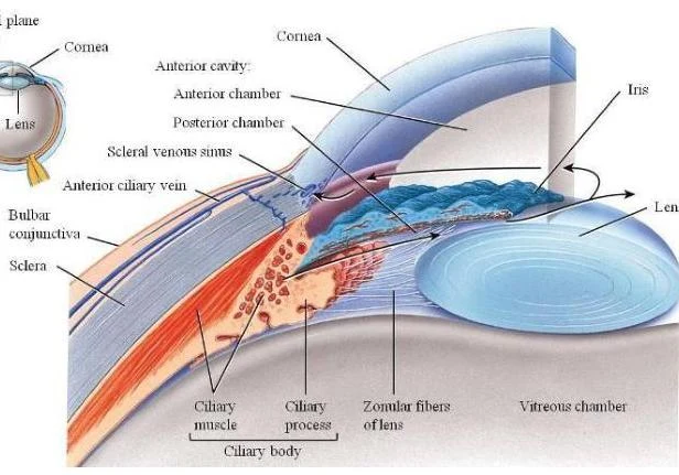
Vascular Tunic (Uvea): The middle layer, comprising the choroid (rich in capillaries), the ciliary body (which secretes aqueous humor and contains the ciliary muscle to alter lens shape), and the iris (the colored part that controls the size of the pupil).
Sensory Tunic (Retina): The innermost layer containing photoreceptors.
Diagram illustrating the anatomy of the human eye, including the cornea, lens, iris, ciliary body, and vitreous chamber.
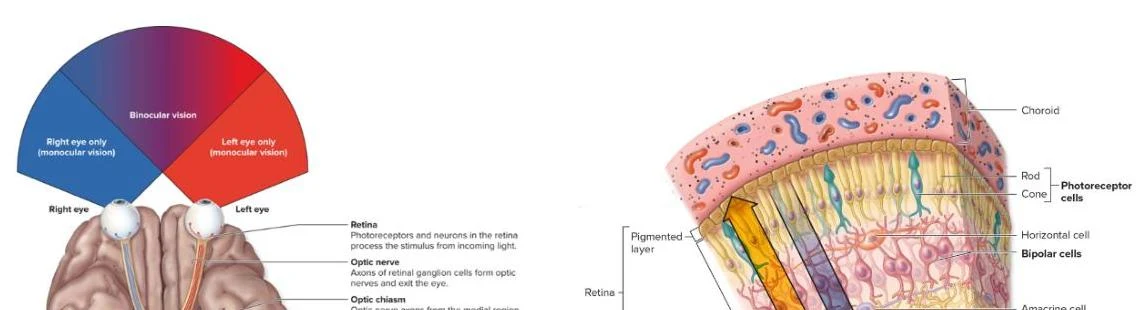
The retina contains two main layers: an outer pigmented layer and an inner neural layer. The neural layer houses photoreceptors: rods for vision in dim light (shades of gray) and cones for bright light and color. The center of the retina features the macula lutea, which contains mostly cones, and the fovea centralis, a region of highest visual acuity containing only cones.
Exam Tip: The Blind Spot
The optic disc is the location where the optic nerve (CN II) exits the eyeball. Because this area lacks photoreceptors, it creates a "blind spot" in our visual field.
The Lens and Internal Cavities
The lens is a transparent, nonvascular structure posterior to the iris that fine-tunes light focusing. The interior of the eye is divided into two cavities by the lens:
Anterior Cavity: Contains the anterior and posterior chambers, filled with aqueous humor which maintains intraocular pressure.
Posterior Cavity (Vitreous Chamber): Filled with a jelly-like vitreous body that presses the retina against the choroid.
Visual Pathway
Sensory information travels from the retina via the optic nerve (CN II), crosses at the optic chiasm, continues as the optic tract, passes through the lateral geniculate body (LGB) of the thalamus, and finally reaches the visual cortex in the occipital lobe.
Diagrams illustrating binocular vision and the structure of the retina, including photoreceptor cells, bipolar cells, and the optic nerve.
The Ear: Audition and Equilibrium
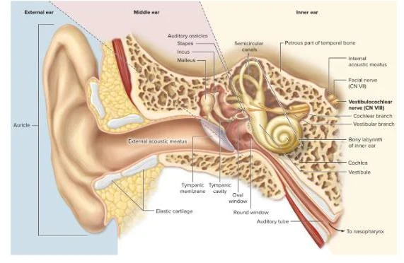
The ear is divided into the external, middle, and internal ear, serving the dual functions of hearing (audition) and balance (equilibrium).
External and Middle Ear
The external ear consists of the auricle (pinna) and the external acoustic canal, which contains ceruminous glands that secrete earwax (cerumen). The canal ends at the tympanic membrane (eardrum).
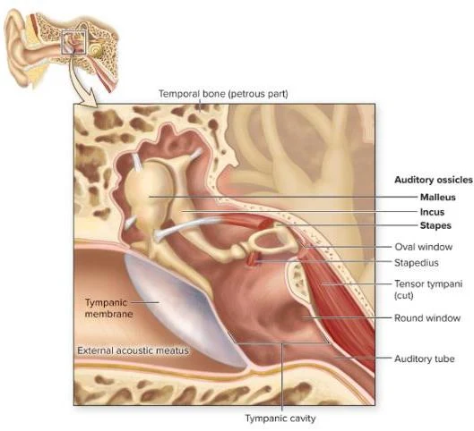
The middle ear is an air-filled cavity containing the three smallest bones in the body, known as ossicles: the malleus, incus, and stapes. The stapes vibrates against the oval window. To protect against loud sounds, the tensor tympani and stapedius muscles can dampen vibrations. The pharyngotympanic (Eustachian) tube connects the middle ear to the nasopharynx to equalize air pressure.
Diagram of the human ear, illustrating the temporal bone, auditory ossicles (malleus, incus, stapes), tympanic membrane, external acoustic meatus, oval window, stapedius muscle, tensor tympani muscle, round window, auditory tube, and tympanic cavity.
Internal Ear: Hearing and Balance
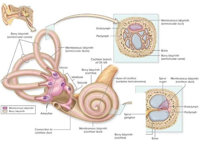
The internal ear contains the bony and membranous labyrinths.
1. Hearing (The Cochlea): The snail-shaped cochlea contains three channels: the scala vestibuli, scala tympani, and the scala media (cochlear duct). The scala media contains the organ of Corti, which houses hair cells that respond to vibrations.
Diagram illustrating the anatomy of the human inner ear, including the bony and membranous labyrinths, semicircular canals, vestibule, saccule, utricle, and cochlea.
2. Equilibrium (Balance):
Static Equilibrium: Monitored by the vestibule (containing the utricle and saccule), which detects linear acceleration and head position relative to gravity.
Dynamic Equilibrium: Monitored by the semicircular canals, which detect rotational or angular acceleration.
The sensory input from both hearing and balance travels via the vestibulocochlear nerve (CN VIII) to the brainstem and eventually to the temporal lobe.
Diagram of the human ear, illustrating the external, middle, and inner ear structures, including the auricle, auditory ossicles, semicircular canals, cochlea, and auditory tube.
Aging and Sensory Health
As humans age, several physiological changes occur in the sensory systems:
Smell and Taste: Deterioration usually becomes noticeable after age 50 as receptor replacement slows down.
Vision: Common issues include presbyopia (loss of lens elasticity), cataracts (loss of lens transparency), glaucoma (high intraocular pressure), and increased "vitreal floaters".
Hearing: Presbycusis (age-related hearing loss, especially for high pitches) is common by age 60.
Medical Terminology Quick Reference
Anosmia: Total loss of smell.
Ageusia: Loss of taste.
Glaucoma: High intraocular pressure that can lead to blindness.
Cataract: Clouding of the lens.
Keep reading for free.
Create an account to finish this reading and make it part of your study routine.