


The thoracic cage is a sophisticated bony framework designed to protect vital organs like the heart and lungs while facilitating the continuous movement required for respiration. This movement is made possible by a series of specialized articulations between the ribs, costal cartilages, sternum, and the vertebral column. Unlike the rigid joints of the skull, the joints of the thorax must balance stability with the flexibility needed to expand and contract the chest during breathing.
Anterior and Lateral Articulations
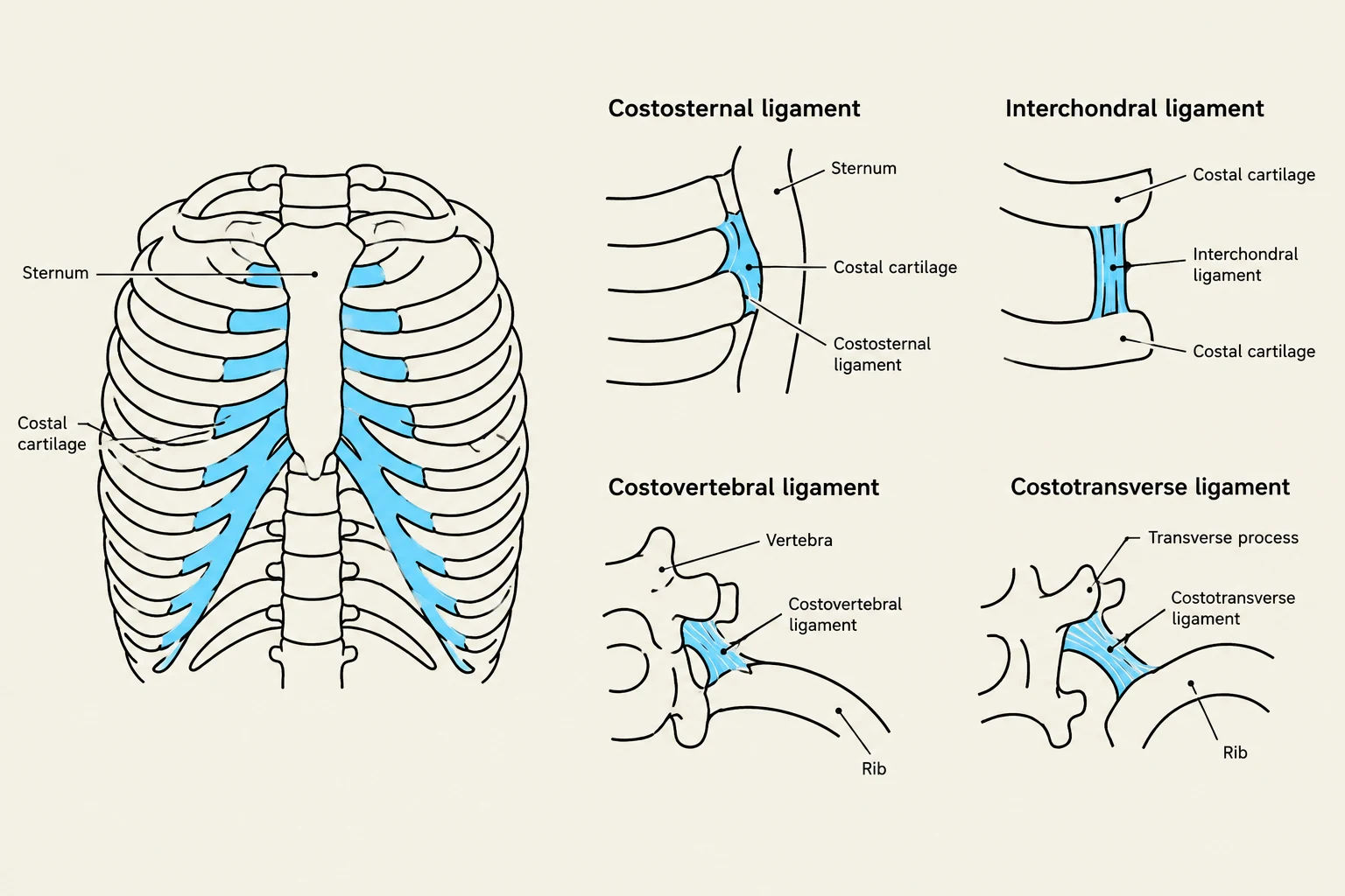
The anterior and lateral aspects of the thorax involve connections between the ribs, their cartilaginous extensions, and the sternum. These joints are categorized by their mobility and the specific tissues involved.
Chondrosternal Joints
The chondrosternal joints connect the costal cartilages of ribs 2 through 7 to the sternum. These are classified as amphiarthrodial joints, meaning they allow for a limited amount of movement. This slight flexibility is essential for the elevation of the ribs during inhalation.
Costochondral Joints
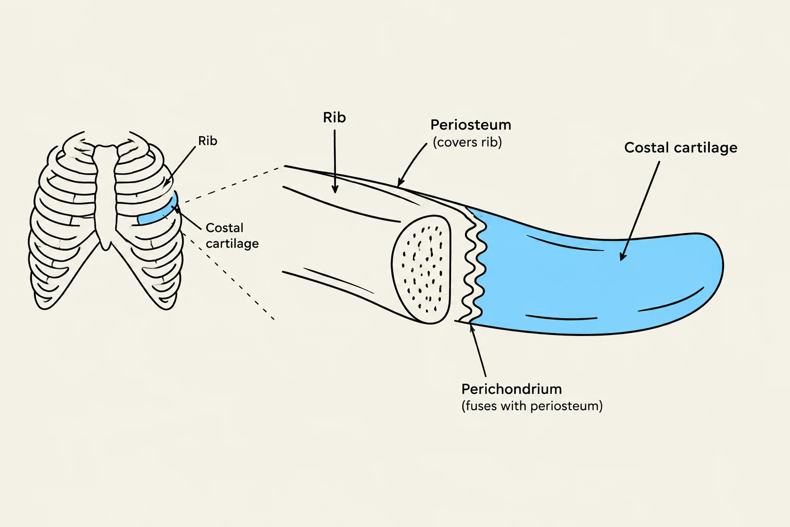
Moving laterally along the rib, the costochondral joints occur at the junction between all ribs and their respective costal cartilages. Interestingly, these joints do not possess specific ligaments to hold them together; instead, the connection is maintained by the direct fusion of the periosteum (the membrane covering the bone) to the perichondrium (the membrane covering the cartilage). Functionally, they act as either synovial or amphiarthrodial joints, facilitating the upward and outward movement of the ribs.

Keep reading for free.
Create an account to finish this reading and make it part of your study routine.
Continue reading freeAlready have an account? Log in
Updated