Explores the anatomy of the thoracic cage, including the sternum and ribs, and its crucial role in respiration, detailing the diaphragm and intercostal muscles.
The human thorax, often referred to as the thoracic cage, serves as a vital protective housing for the body's most essential organs. This bony frame is not a static shield; rather, it is a dynamic structure designed to facilitate the continuous process of respiration. By balancing rigidity for protection with flexibility for movement, the thorax ensures that the heart, lungs, and major vessels are both safe and functional.
Composition and Function of the Thoracic Cage
The thoracic cage is a complex anatomical arrangement composed of three primary skeletal components:
Thoracic Vertebrae: Located posteriorly, these twelve vertebrae provide the structural foundation and articulation points for the ribs.
Ribs: Located laterally, these twelve pairs of bones form the sides of the cage.
Sternum: Located anteriorly, this "breastbone" completes the front of the cage.
The functions of this cage are multifaceted. Primarily, it provides a protective enclosure for the heart, lungs, trachea, and esophagus. Beyond protection, the thorax serves as a critical attachment point for the pectoral girdle and the upper limbs. Finally, its ability to expand and contract is the mechanical basis for respiration.
While the thorax provides stability, the trunk associated with this region is capable of significant movement, including:
Flexion: ≈80∘
Extension: 20–30∘
Rotation: ≈45∘
Lateral Flexion: ≈35∘
Diagram of the human rib cage, highlighting the thoracic vertebrae, ribs, and sternum.
The Sternum: Regions and Landmarks
The sternum is a flat bone located in the midline of the anterior chest. It is divided into three distinct anatomical regions, each with specific roles and landmarks:
The Manubrium
The manubrium is the most superior portion of the sternum. It features two important landmarks:
Suprasternal notch: A palpable indentation located at the base of the throat.
Clavicular notches: Specialized areas on either side that allow for articulation with the clavicles (collarbones).
The Body
The body of the sternum is the longest, central segment of the bone. It serves as a major attachment site for the costal cartilages of the middle ribs.
The Xiphoid Process
The xiphoid process is the smallest, most inferior tip of the sternum. While it may seem insignificant, it provides essential attachment points for several abdominal muscles, such as the rectus abdominis.
Rib Anatomy and Classification
The ribs are organized into twelve pairs, and they are classified based on how they attach to the sternum.
Rib Classifications
True Ribs (1–7): These seven pairs are considered "true" because each possesses its own dedicated costal cartilage that attaches directly to the sternum.
False Ribs (8–12): This group consists of five pairs. Ribs 8–10 are considered false because they do not attach directly to the sternum; instead, they attach indirectly to the costal cartilage of the 7th rib.
Floating Ribs (11–12): A specific subset of false ribs, these two pairs have no anterior attachment to the sternum at all. Instead, they are embedded within the surrounding musculature.
Structural Landmarks
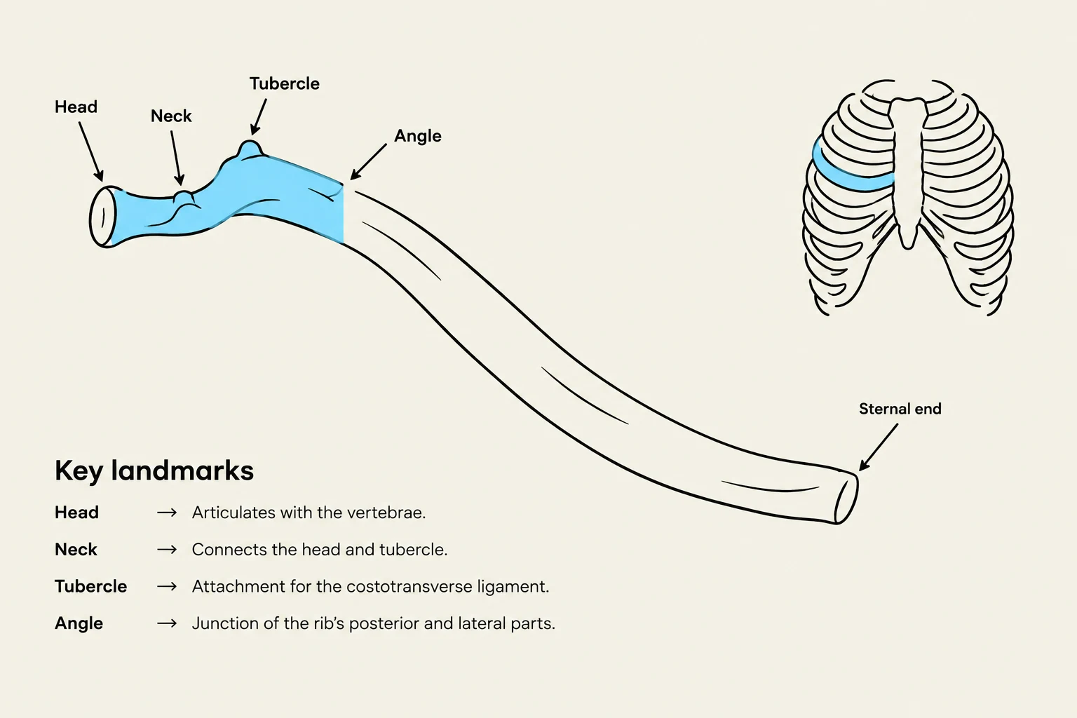
Each rib contains specific features that allow it to articulate with the thoracic vertebrae. These include the head, the neck, the articular facet, the tubercle, and the angle. The angle of the rib is particularly important for posture; it contributes to a broad back structure that allows the body to lie supine (on the back) without rolling to the side.
Diagram illustrating the key landmarks of a rib bone, including the head, neck, tubercle, angle, and sternal end, with descriptions of their functions.
Respiratory Mechanics: The Muscles of the Thorax
Respiration is driven by changes in the volume and pressure of the thoracic cavity, which are controlled by specific muscles.
The Diaphragm
The diaphragm is the primary muscle of respiration. It is a broad, dome-shaped muscle that separates the thoracic cavity from the abdominal cavity.
Origin (O): It originates from the xiphoid process, the costal cartilages of ribs 6–12, and the lumbar vertebrae (L1–L4).
Insertion (I): It inserts into the central tendon, a wide aponeurosis that forms the top of the diaphragm and blends with the fibrous pericardium.
Action (A): During inspiration, the diaphragm contracts and the central tendon pulls downward. This flattens the muscle, increasing the volume of the thoracic cavity and lowering internal pressure to draw air in.
Clinical Note: The diaphragm is a vital passageway for major structures. The inferior vena cava passes through the central tendon, while the aorta and esophagus also pierce the muscle to allow passage between the thorax and abdomen.
Intercostal Muscles
The muscles located between the ribs, known as intercostals, assist in regulating the movement of the rib cage:
External Intercostals:
Origin: Inferior border of the rib above.
Insertion: Superior border of the rib below.
Action: They elevate the ribs, which helps expand the chest during inspiration.
Internal Intercostals:
Origin: Inferior border (specifically the costal groove) of the rib above.
Insertion: Superior border of the rib below.
Action: They depress the ribs, compressing the thoracic cavity to assist in forced expiration.
Diagram illustrating the orientation and fiber directions of external and internal intercostal muscles in the rib cage.
Summary Table: Respiratory Muscle Comparison
Muscle
Primary Action
Phase of Respiration
Diaphragm
Flattens/Pulls down
Inspiration (Primary)
External Intercostals
Elevates ribs
Inspiration (Assisted)
Internal Intercostals
Depresses ribs
Forced Expiration
Keep reading for free.
Create an account to finish this reading and make it part of your study routine.