


The human foot is a sophisticated mechanical structure designed to balance two conflicting requirements: providing a stable platform for weight-bearing and offering the flexibility necessary for locomotion. To achieve this, the foot utilizes a complex arrangement of 26 bones, organized into functional groups, and is powered by muscles controlled by a highly organized neural network. Understanding the relationship between these osseous structures and their innervation is fundamental to clinical assessment, particularly when evaluating gait abnormalities or nerve injuries.
The Osseous Architecture of the Foot
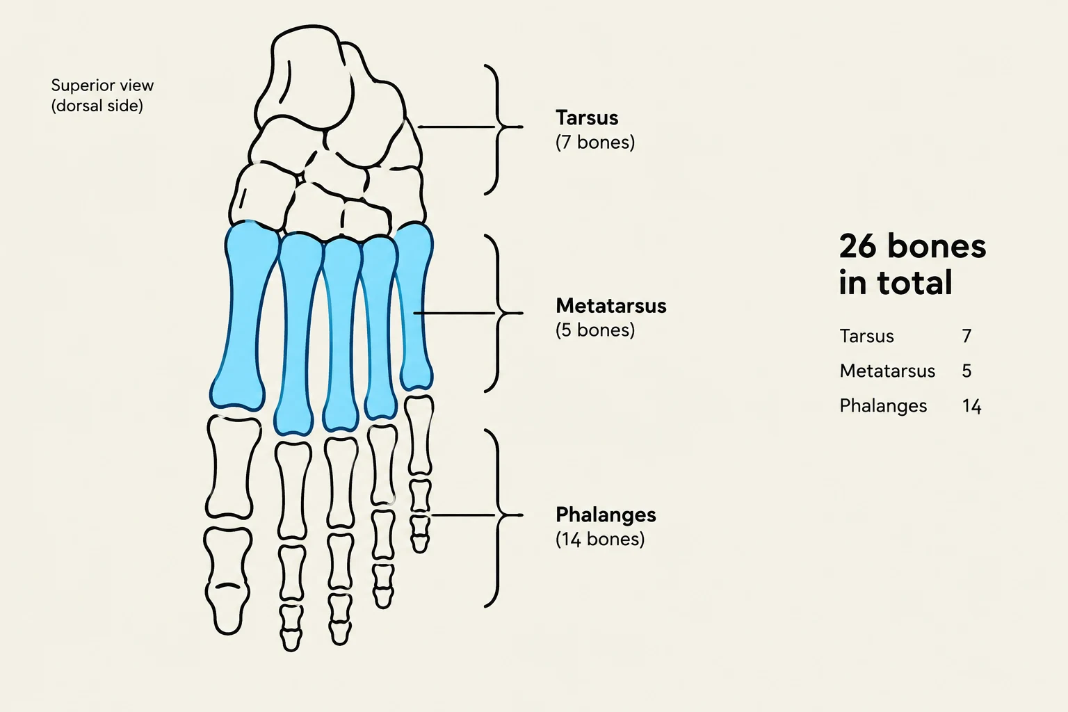
The Tarsus (Hindfoot and Midfoot)
The tarsus consists of seven bones that form the foundation of the foot, providing the structural integrity needed to support body weight.
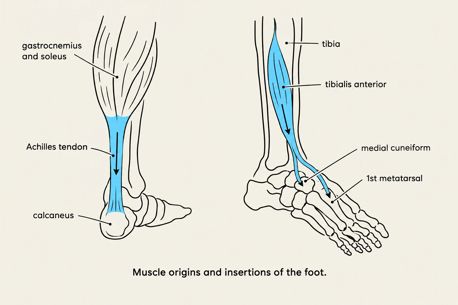
- Calcaneus: This is the largest of the tarsal bones and forms the heel. It serves as a critical lever arm for the Achilles tendon, facilitating powerful plantarflexion.
- Talus: Positioned atop the calcaneus, the talus acts as the "keystone" of the foot. It articulates with the tibia and fibula to form the ankle mortise.
- Navicular: A boat-shaped bone located on the medial side, situated between the talus and the cuneiforms.
- Cuboid: Located on the lateral aspect of the foot, it articulates with the calcaneus proximally and the fourth and fifth metatarsals distally.
- Cuneiforms: There are three wedge-shaped cuneiform bones—medial, intermediate, and lateral. They articulate with the navicular posteriorly and the first three metatarsals anteriorly.

Keep reading for free.
Create an account to finish this reading and make it part of your study routine.
Continue reading freeAlready have an account? Log in
Updated