


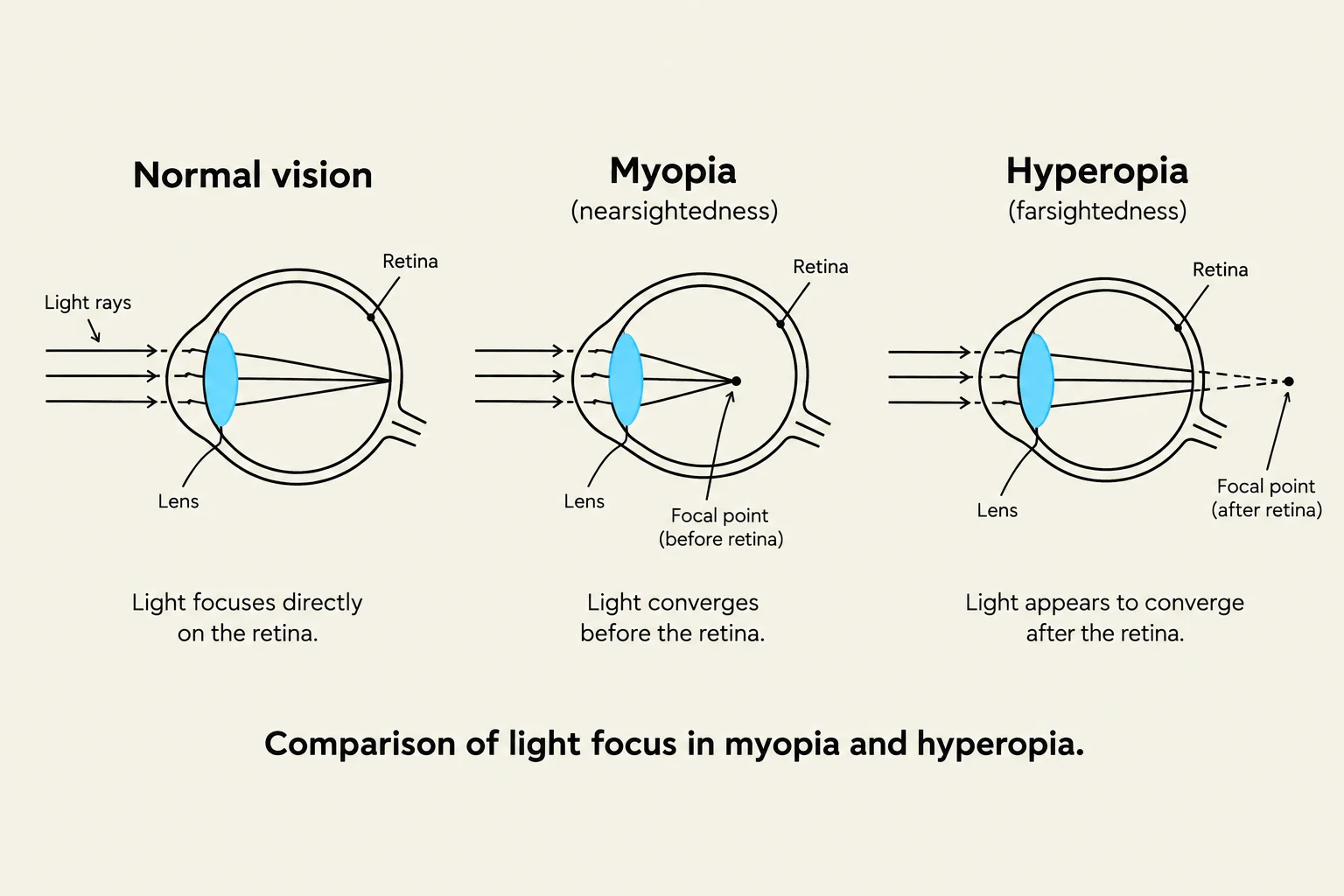
Vision is a complex psychological and biological process that begins with sensation—the detection of physical stimuli by sensory receptors—and culminates in perception, where the brain organizes and interprets that information to create a meaningful experience. While your eyes detect light (sensation), it is your brain that identifies that light as a "red rose" (perception). This article explores the intricate anatomy of the eye, the mechanism of light transduction, and the common visual impairments that affect how we see the world.
The Structure of the Eye
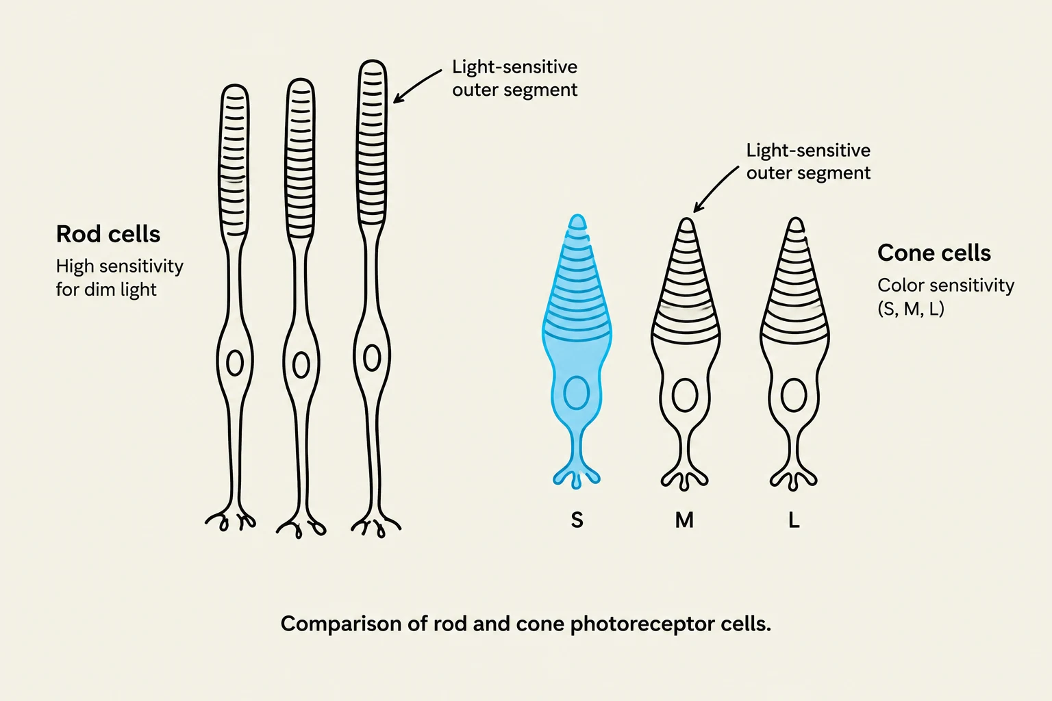
The human eye acts as a sophisticated biological camera, designed to collect and focus light so that it can be converted into neural signals. This conversion process is known as transduction.
The Outer and Middle Layers: Cornea, Iris, and Pupil
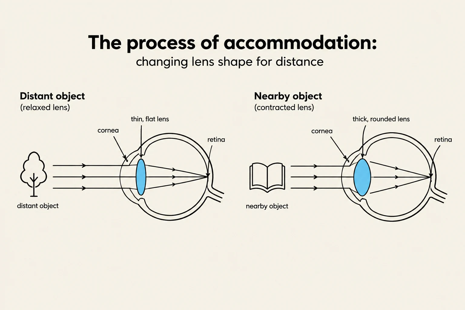
The journey of light begins at the cornea, a transparent outer surface that serves the critical function of refracting (bending) incoming light. Once light passes through the cornea, it reaches the iris, the colored muscular structure of the eye.
The iris does not just provide color; it acts as a regulator for the pupil, which is the opening in the center of the iris. By adjusting the diameter of the pupil, the iris controls the amount of light entering the eye:
- In bright light: The pupil constricts (becomes smaller) to prevent overexposure.
- In dim light: The pupil dilates (becomes larger) to allow more light to reach the retina.
Exam Tip: Remember that the pupil itself is not a muscle; it is an opening. The iris is the muscular structure that performs the action of changing the pupil's size.
Keep reading for free.
Create an account to finish this reading and make it part of your study routine.
Continue reading freeAlready have an account? Log in
Updated