Anatomy and Functions of the Mouth, Pharynx, and Esophagus in Digestion
This article explores the initial stages of the digestive process, detailing the anatomy and functions of the mouth, pharynx, and esophagus, including mechanical and chemical digestion, saliva production, and the process of swallowing (deglutition).
The digestive process is a complex sequence of activities designed to transform food into absorbable nutrients. This journey begins with ingestion, the entry of food into the alimentary canal through the mouth. Before nutrients can reach the bloodstream, food must undergo mechanical breakdown and chemical digestion to increase its surface area and reduce complex molecules into their building blocks. This article explores the first critical stages of this process: the mouth, the pharynx, and the esophagus.
The Mouth: The Gateway to Digestion
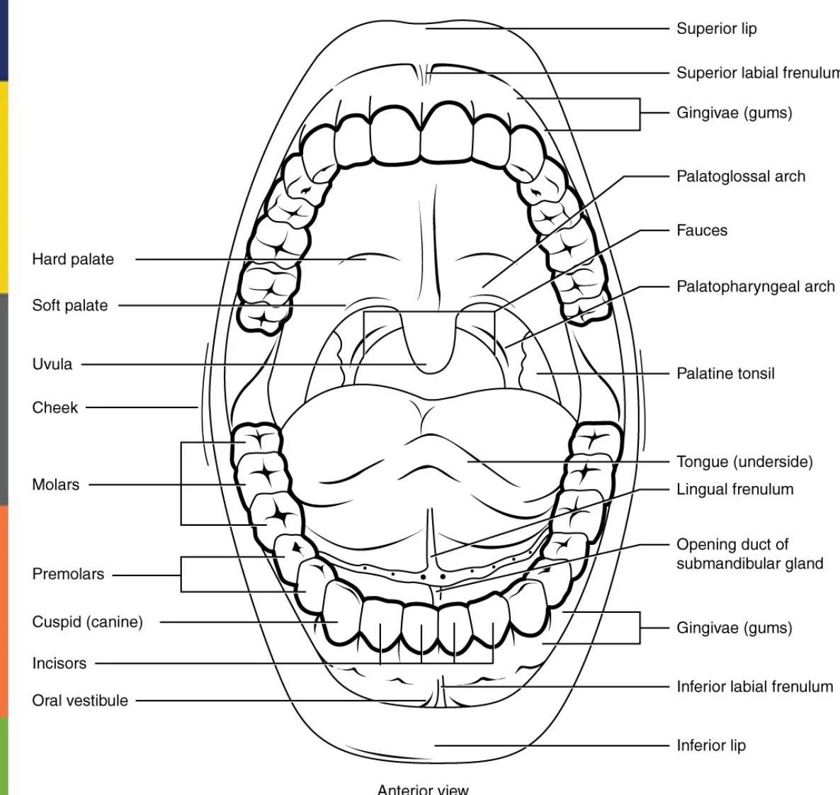
The mouth is the primary site for both ingestion and the beginning of mechanical and chemical digestion. It is composed of several specialized structures, including the lips (labia), cheeks, tongue, palate, gums, and teeth.
An anterior view diagram illustrating the anatomy of the human mouth, labeling various structures such as the lips, tongue, teeth, palate, uvula, and glands.
Mastication and Mechanical Digestion
Mastication, or chewing, is a form of mechanical digestion. During this process, the teeth shred and crush food into smaller particles, while the lips and cheeks work to confine the food between the teeth to ensure even chewing. This physical breakdown is essential because it increases the surface area of the food, making it more accessible to digestive enzymes.
The Role of the Tongue and Teeth
The tongue is a versatile organ that facilitates ingestion, sensation, and swallowing. Its functions are divided between two muscle groups:
Extrinsic muscles: Move the tongue sideways, in, and out to manipulate food for chewing and to shape it into a bolus (a soft mass of chewed food).
Intrinsic muscles: Change the shape of the tongue to assist in swallowing.
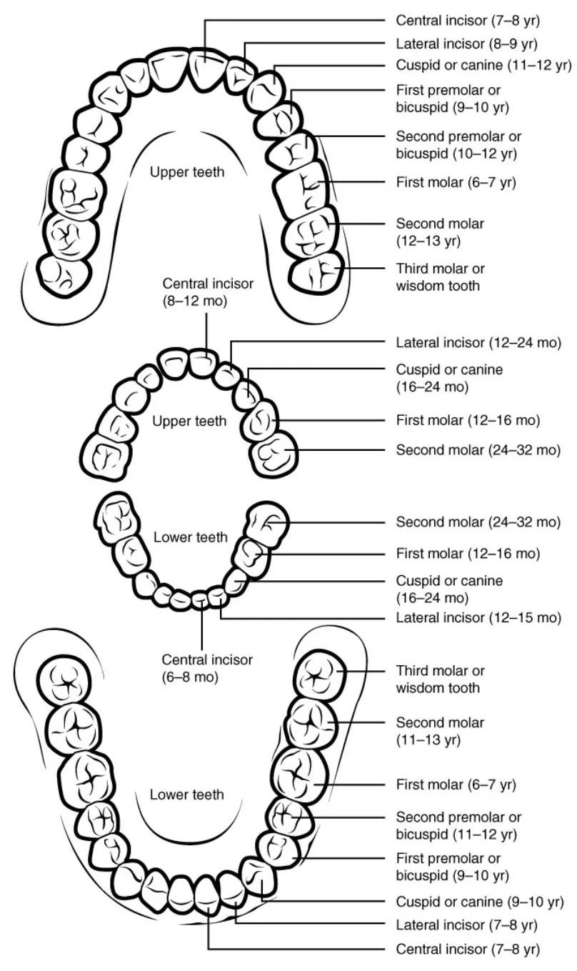
The teeth are responsible for the physical destruction of solid food. Humans transition from a set of 20 deciduous teeth (baby teeth) to a set of 32 permanent teeth.
Diagrams illustrating the eruption timelines of deciduous and permanent teeth in children.
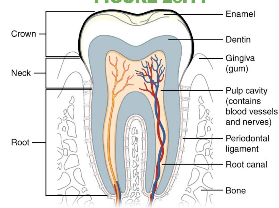
The structure of a tooth (dens) includes several distinct layers:
Enamel: The hardest substance in the human body, composed mostly of calcium salts; it cannot be replaced if damaged.
Dentin: A layer similar to bone but harder than dentin.
Pulp Cavity: The innermost part containing nerves, blood vessels, and connective tissue.
Periodontal Ligament: Attaches the tooth to the jaw.
Diagram illustrating the anatomy of a tooth, including enamel, dentin, pulp cavity, root canal, and surrounding structures like the gingiva and bone.
Saliva and Chemical Digestion
While mastication handles the physical aspect, chemical digestion begins immediately in the mouth through the secretion of saliva. Saliva is approximately 95.5% water and contains mucus, which moistens and dissolves food to allow for taste.
Salivary Glands and Enzymes
Major salivary glands are located outside the oral mucosa and deliver saliva through ducts. These glands secrete enzymes that begin the chemical breakdown of macronutrients:
Salivary amylase: Breaks down polysaccharides (carbohydrates).
Lingual lipase: Secreted by lingual glands, it begins the breakdown of triglycerides (fats).
Enzyme Category
Enzyme Name
Source
Substrate
Salivary Enzymes
Lingual lipase
Lingual glands
Triglycerides
Salivary Enzymes
Salivary amylase
Salivary glands
Polysaccharides
Regulation of Salivation
Salivation is regulated by the autonomic nervous system:
Parasympathetic Nervous System: Provides constant low-level stimulation and can trigger large amounts of saliva in response to sensory stimulation or the thought of food.
Sympathetic Nervous System: Activation results in small amounts of saliva, often leading to a "dry mouth" sensation.
Exam Tip: Remember that taste buds do more than just sense flavor; they send nerve impulses to the salivary nuclei in the brain stem, which then trigger the salivary glands to secrete saliva.
The Pharynx and Esophagus: Propulsion
Once the food is prepared as a bolus, it must be moved toward the stomach through the processes of propulsion.
The Pharynx
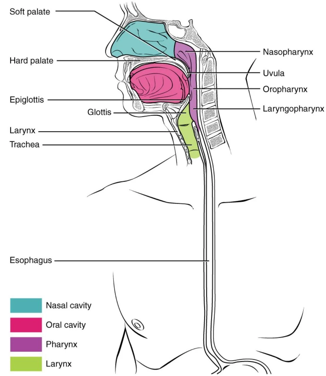
The pharynx is a tube of skeletal muscle lined with a mucous membrane that serves both the digestive and respiratory systems. It runs from the nostrils to the esophagus and the larynx.
Diagram of the human upper airway and digestive tract, illustrating the nasal cavity, oral cavity, pharynx, larynx, trachea, and esophagus.
The Esophagus and Peristalsis
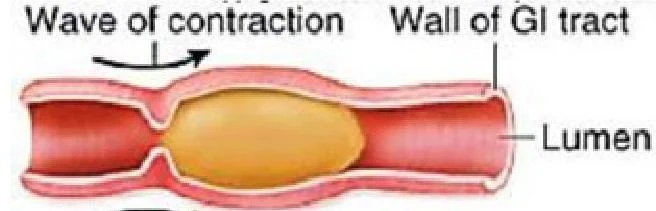
The esophagus is a muscular tube that propels food to the stomach. This movement is achieved through peristalsis, a series of alternating waves of contraction and relaxation of the smooth muscles in the alimentary wall. Peristalsis is powerful enough to move food to the stomach even if an individual is upside down.
Diagram illustrating peristalsis, the wave of muscular contractions that moves food through the gastrointestinal tract.
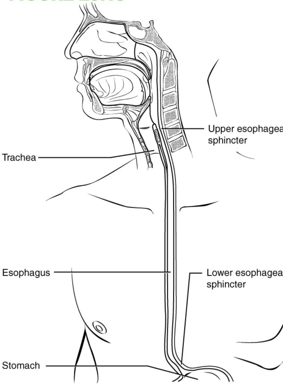
To ensure food moves in the correct direction and does not reflux, the esophagus utilizes two sphincters:
Upper Esophageal Sphincter: Controls the movement of food from the pharynx into the esophagus.
Lower Esophageal Sphincter: Allows the bolus to enter the stomach and prevents acidic chyme from entering the esophagus.
Diagram of the human digestive and respiratory tracts, showing the trachea, esophagus, upper and lower esophageal sphincters, and stomach.
Clinical Note: The esophagus penetrates the diaphragm through an opening called the esophageal hiatus. If the stomach pushes through this opening, it results in a sliding hiatal hernia.
Deglutition (Swallowing)
Deglutition is the process of swallowing and represents the last voluntary act in the digestive process before the movement becomes entirely involuntary. It consists of three distinct phases:
Voluntary Phase: The initial act of moving the bolus toward the back of the mouth.
Pharyngeal Phase: An involuntary phase where the bolus is propelled from the pharynx into the esophagus.
Esophageal Phase: An involuntary phase where peristalsis moves the bolus through the esophagus to the stomach.
Keep reading for free.
Create an account to finish this reading and make it part of your study routine.