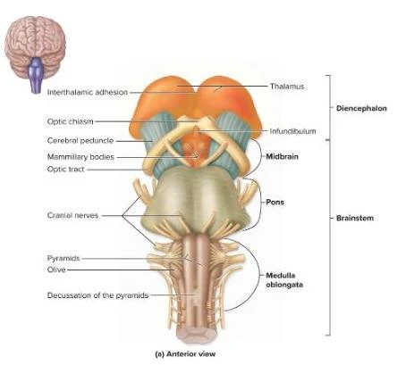
1. Midbrain (Mesencephalon)
The midbrain is the superior portion of the brainstem. A key feature is the cerebral aqueduct, which allows cerebrospinal fluid to flow from the third ventricle to the fourth ventricle. The posterior portion, known as the tectum, contains two important structures:
- Superior Colliculi: These manage reflex movements of the eyes, head, and neck.
- Inferior Colliculi: These serve as a relay station for auditory information.
2. Pons
The pons is the bulging middle region of the brainstem. It serves two primary functions:
- Respiratory Regulation: It contains the pontine respiratory center, which helps regulate the rhythm of breathing.
- Auditory Processing: Through the superior olivary complex, it receives auditory input to help localize where a sound is coming from.
3. Medulla Oblongata
The medulla is the most inferior part of the brainstem, transitioning directly into the spinal cord. It is perhaps the most critical area for immediate survival, as it contains:
- Vital Autonomic Centers: These include the cardiovascular center (heart rate/blood pressure), the medullary respiratory center (breathing), and the vasomotor center (blood vessel constriction/dilation).
- Reflex Centers: It manages involuntary reflexes such as vomiting, coughing, and sneezing.
The Cerebellum: Coordination and Balance
Located posterior to the brainstem, the cerebellum is the second-largest part of the brain. While it does not initiate movement, it is responsible for the "quality" of that movement.
The cerebellum is structured into three layers:
- An outer layer of gray matter called the cerebellar cortex.
- An internal region of white matter known as the arbor vitae (meaning "tree of life").
- Deep cerebellar nuclei.
Its primary functions include coordinating and fine-tuning motor movements, maintaining equilibrium and posture, and storing motor memories (such as the "muscle memory" used when riding a bike).
The Limbic System: The Emotional Brain
The limbic system is not a single structure but a ring of cerebral and diencephalic structures that surround the diencephalon. It is the seat of our emotional experiences. Key components include the hippocampus (crucial for memory) and the amygdaloid body (involved in emotional expression). This system allows us to process and experience complex emotions and is closely linked to the formation of long-term memories.
Cranial Nerves: The Direct Links to the Body
Cranial nerves are 12 pairs of nerves that emerge directly from the brain rather than the spinal cord. They are classified into three categories based on the type of fibers they carry: Special Sensory, Motor, or Mixed.
Special Sensory Nerves
These nerves carry only sensory information from the environment to the brain:
- I. Olfactory Nerve: Responsible for the sense of smell.
- II. Optic Nerve: Responsible for the sense of sight.
- VIII. Vestibulocochlear Nerve: Responsible for hearing and equilibrium (balance).
Motor Nerves
These nerves carry only motor commands from the brain to the muscles:
- III. Oculomotor Nerve: Controls most eye movements.
- IV. Trochlear Nerve: Controls downward eye movement.
- VI. Abducens Nerve: Controls lateral (side-to-side) eye movement.
- XI. Accessory Nerve: Controls head and neck movement.
- XII. Hypoglossal Nerve: Controls the muscles of the tongue for speech and swallowing.
Mixed Nerves
These nerves are versatile, carrying both sensory and motor fibers:
- V. Trigeminal Nerve: Provides sensation to the face and controls the muscles used for chewing.
- VII. Facial Nerve: Controls facial expressions and provides the sense of taste to the anterior two-thirds of the tongue.
- IX. Glossopharyngeal Nerve: Involved in swallowing and provides taste to the posterior one-third of the tongue.
- X. Vagus Nerve: A massive nerve that travels to the viscera (internal organs), controlling visceral muscle movement, speech, and sensation.
💡 Exam Tip: The Mnemonic
To remember the order of the nerves (I through XII), many students use a mnemonic. A common one is: "Oh, Oh, Oh, To Touch And Feel Very Good Velvet, AH!" where each word represents the starting letter of the nerve name.
| Nerve Type | Nerve Numbers | Primary Function |
|---|
| Sensory | I, II, VIII | Sensation (Smell, Sight, Hearing) |
| Motor | III, IV, VI, XI, XII | Movement (Eyes, Neck, Tongue) |
| Mixed | V, VII, IX, X | Both Sensation and Movement |
References
McKinley Anat 32 Ch 15.docx.pdf