


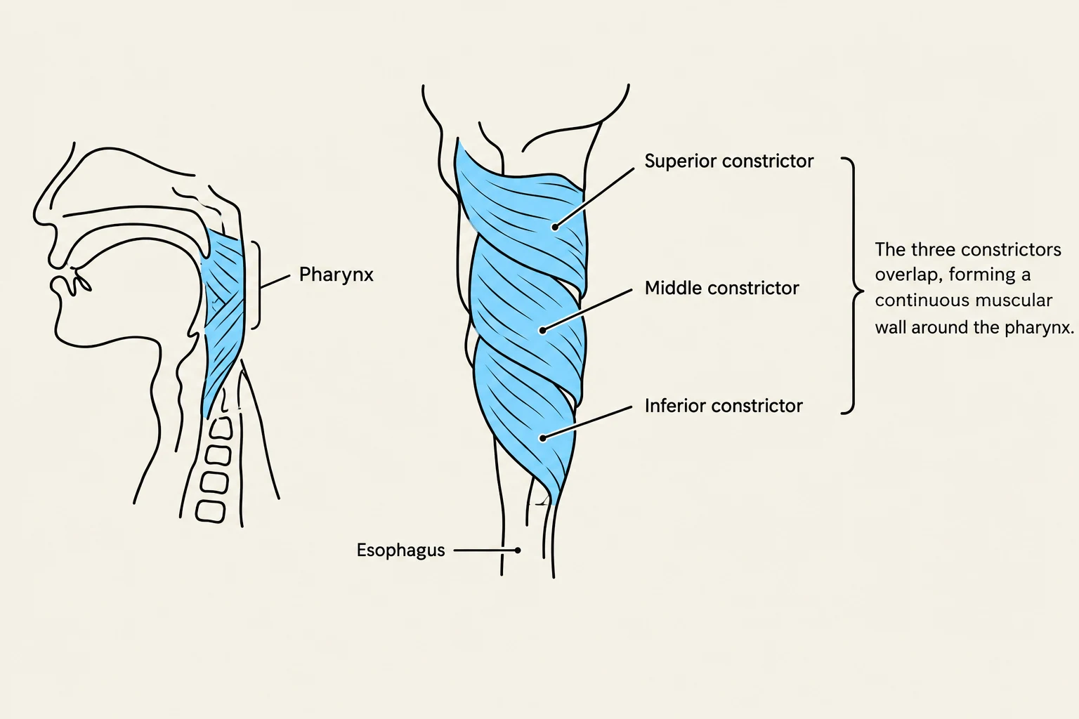
The soft palate and the pharynx are complex muscular structures that work in perfect coordination to facilitate essential life functions: breathing, speaking, and swallowing. While the soft palate forms the posterior end of the roof of the mouth, the pharynx serves as a muscular tube divided into three distinct regions: the nasal pharynx, the oral pharynx, and the laryngeal pharynx. Understanding the specific origins, insertions, and nerve supplies of these muscles is key to understanding how we transition food from the mouth to the esophagus without entering the airway.
The Soft Palate
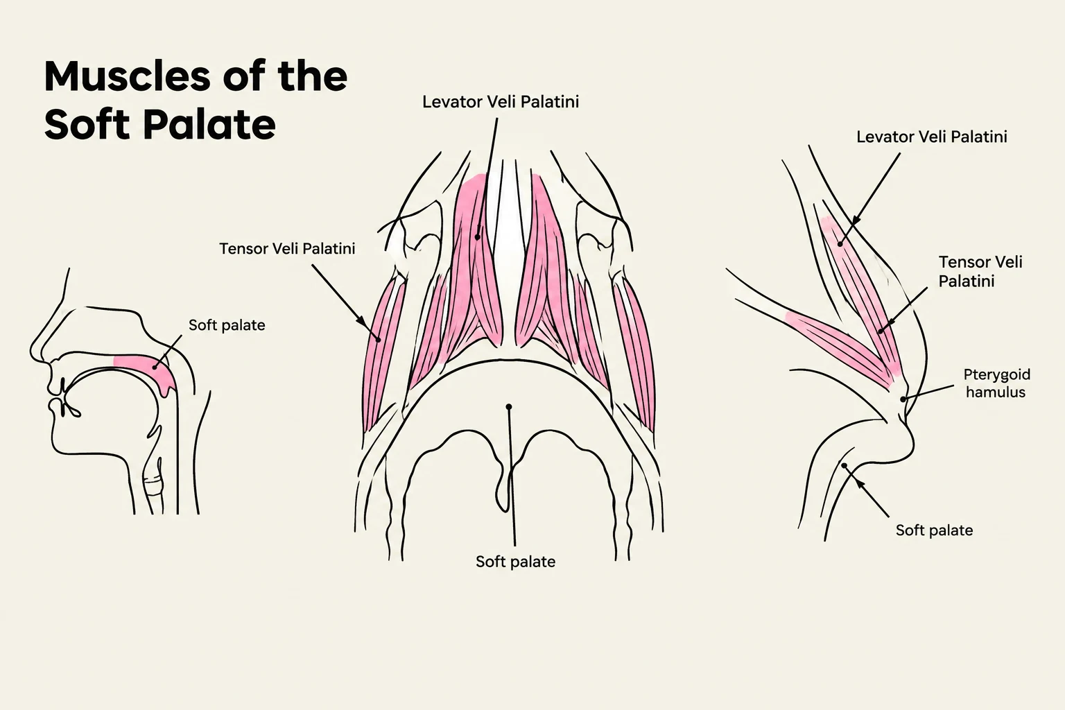
The soft palate consists of five pairs of muscles that work together to move the palate up and back. This movement allows the palate to contact the posterior throat wall, effectively sealing off the nasal cavity and narrowing the fauces—the space located between the palatine tonsils.
Muscles of the Soft Palate
Several specific muscles govern the movement and tension of this region:
- Palatoglossus Muscle: Located under the palatoglossal fold (the anterior faucial pillar), this muscle originates from the posterior end of the hard palate and the anterior end of the soft palate. It runs downward and laterally to insert into the posterolateral part of the tongue. Its contraction pulls the soft palate down on its lateral edges, narrowing the space between the anterior faucial pillars.
- Palatopharyngeal Muscle: This muscle forms the posterior faucial pillar. It originates from the posterolateral part of the soft palate and inserts into the pharyngeal constrictor muscle and the thyroid cartilage of the larynx. When it contracts, it narrows the posterior faucial pillar and elevates both the pharynx and the larynx.
- Levator Veli Palatini: This muscle is responsible for elevating the posterior end of the soft palate. It originates from the petrous part of the temporal bone and the medial wall of the auditory canal. Its contraction pulls the soft palate up and back against the pharyngeal wall and can also open the auditory tube.
- Tensor Veli Palatini: Originating near the base of the medial pterygoid plate and the lateral wall of the auditory tube, this muscle inserts into the posterior edge of the hard palate and the anterior portion of the soft palate. It functions to tense the anterior portion of the soft palate and open the auditory tube.
- Muscles of the Uvula: The uvula is a small fold of tissue hanging from the posterior soft palate. It is formed by two small bands of muscle that originate from the posterior end of the hard palate. Contraction shortens and broadens the uvula, helping the soft palate adapt to the contour of the posterior throat wall.

Keep reading for free.
Create an account to finish this reading and make it part of your study routine.
Continue reading freeAlready have an account? Log in
Updated