


Understanding the musculature of the pelvis and hip is essential for grasping how the human body achieves both powerful movement and intricate stability. This region serves as the primary link between the axial skeleton (the spine) and the lower limbs, facilitating everything from high-impact locomotion to the subtle postural adjustments required for sitting and standing.
Pelvic Landmarks and Osteology
Before palpating muscles, a clinician must be able to identify the bony landmarks that serve as attachment points. The pelvis is a complex structure, and its surface anatomy provides critical cues for locating underlying musculature.
Key landmarks include:
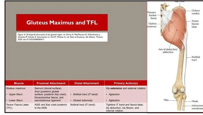
- Posterior Iliac Crest: The superior border of the ilium, which is easily palpable at the waistline.
- Anterior Superior Iliac Spine (ASIS): A prominent bony projection at the front of the pelvis.
- Sacrum and Coccyx: The base of the vertebral column that integrates into the pelvic ring.
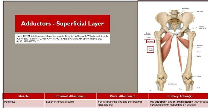
- Femur: The thigh bone, specifically the greater trochanter and the femoral shaft, which serves as a major distal attachment site for hip muscles.
Muscle Groups: Attachments and Actions
The muscles of the pelvis and hip can be categorized by their location and their primary contribution to joint movement. Each muscle has a proximal attachment (origin) and a distal attachment (insertion).
1. Hip Flexors
These muscles are located primarily on the anterior aspect of the hip and are responsible for bringing the thigh toward the abdomen.
- Primary Action: Hip flexion.
- Key Muscles: Includes the Iliopsoas and Tensor Fasciae Latae (TFL).
Keep reading for free.
Create an account to finish this reading and make it part of your study routine.
Continue reading freeAlready have an account? Log in
Updated