Anatomy and Function of the Central Nervous System
An overview of the central nervous system's structure, including the brain, brainstem, cerebellum, and ventricles. It details the organization of gray and white matter, lobes of the cerebrum, and the functions of key regions like the diencephalon.
The Central Nervous System (CNS) is the command center of the human body, responsible for processing sensory information, initiating voluntary movement, and managing complex cognitive functions like memory and emotion. Understanding its anatomy is crucial for clinical practice, particularly when addressing injuries such as strokes or degenerative diseases. This article explores the structural organization, tissue composition, and functional regions of the adult CNS.
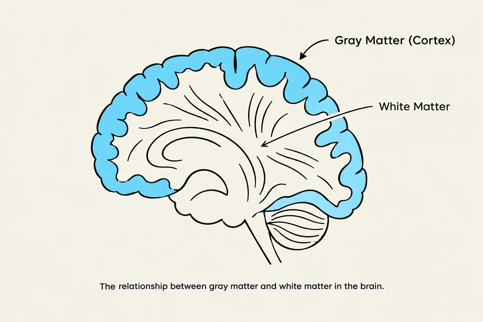
The relationship between gray matter and white matter in the brain.
Structural Organization and Tissue Composition
The adult brain is organized into four primary regions: the cerebral hemispheres, the diencephalon, the brain stem (which includes the midbrain, pons, and medulla oblongata), and the cerebellum.
While the spinal cord follows a consistent pattern of a central gray matter cavity surrounded by external white matter, this distribution shifts as we move into the brain. The CNS is composed of two distinct types of tissue:
Gray matter: Consists of short, nonmyelinated neurons and cell bodies.
White matter: Consists of myelinated and nonmyelinated axons.
In the cerebral hemispheres and the cerebellum, the gray matter forms an outer layer known as the cortex. Beneath this cortex lies the internal white matter, which contains scattered nuclei of gray matter.
The Cerebral Hemispheres
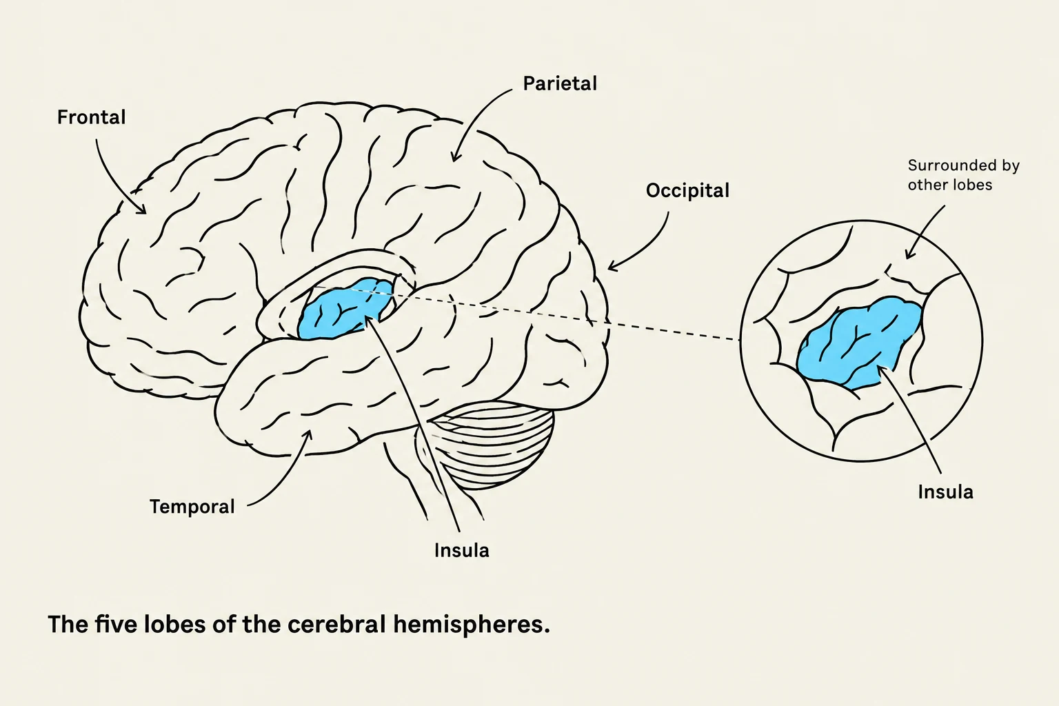
Diagram illustrating the five lobes of the cerebral hemispheres: frontal, parietal, temporal, occipital, and insula.
The cerebral hemispheres constitute the largest portion of the brain, accounting for approximately 83% of its total mass. Their surface is not smooth; instead, it is characterized by various folds that increase surface area:
Gyri: The ridges or bumps on the surface.
Sulci: Shallow grooves between the ridges.
Fissures: Deep grooves that separate major regions.
Two major landmarks are the longitudinal fissure, which separates the left and right hemispheres, and the transverse cerebral fissure, which separates the cerebrum from the cerebellum.
Lobes of the Cerebrum
The hemispheres are divided into five distinct lobes by various sulci:
Frontal lobe: Involved in motor control and higher-level functions.
Parietal lobe: Primarily associated with sensory perception.
Temporal lobe: Involved in auditory processing and memory.
Occipital lobe: The center for visual processing.
Insula: A lobe buried deep beneath the temporal, parietal, and frontal lobes.
The Cerebral Cortex: The "Executive Suite"
The cerebral cortex is a thin (2–4 mm) superficial layer of gray matter that serves as the site of the conscious mind. It is responsible for sensory perception, voluntary motor initiation, communication, and memory storage. Functional imaging (such as fMRI) shows that specific functions are located in discrete cortical areas called domains.
The cortex contains three types of functional areas:
Motor areas: Control voluntary movement.
Sensory areas: Provide conscious awareness of sensation.
Association areas: Integrate diverse information to provide meaning.
Exam Tip: Lateralization
Remember that the two hemispheres are not identical. This is called lateralization. In about 90% of humans, the left hemisphere is dominant for language, math, and logic, while the right hemisphere specializes in visual-spatial skills, intuition, and emotion.
The Diencephalon
The diencephalon is located deep within the brain and consists of three paired gray matter structures that enclose the third ventricle:
Thalamus: Making up 80% of the diencephalon, it acts as a relay station. It sorts, edits, and relays ascending sensory impulses and motor impulses to the appropriate cortical areas.
Hypothalamus: Located below the thalamus, it is the main visceral control and regulating center vital to homeostasis. It regulates the autonomic nervous system, body temperature, hunger, thirst, and the endocrine system via the pituitary gland.
Epithalamus: The most dorsal portion, which contains the pineal gland that secretes melatonin to regulate sleep-wake cycles.
The Brain Stem and Cerebellum
The Brain Stem
The brain stem serves as a conduit for fiber tracts and contains nuclei associated with 10 of the 12 pairs of cranial nerves. It consists of three regions:
Midbrain: Located between the diencephalon and pons; it contains the cerebral aqueduct and structures like the substantia nigra.
Pons: Located between the midbrain and medulla; it helps maintain the rhythm of breathing and relays impulses between the motor cortex and cerebellum.
Medulla Oblongata: The most inferior part, blending into the spinal cord. It acts as an autonomic reflex center, regulating cardiovascular and respiratory functions.
The Cerebellum
Accounting for about 11% of brain mass, the cerebellum is located dorsal to the pons and medulla. Its primary role is to provide precise, coordinated movements of skeletal muscles and to maintain balance. It functions by receiving "intent" from the cerebral cortex and "positional" data from proprioceptors, then calculating a "blueprint" for smooth movement.
Ventricles and Cerebrospinal Fluid (CSF)
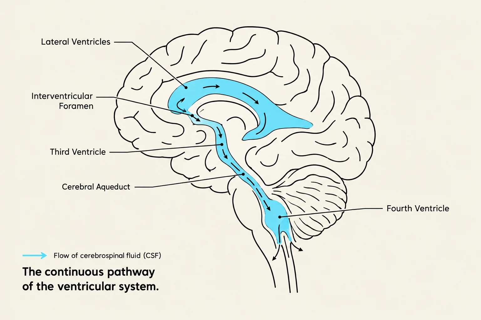
Diagram illustrating the flow of cerebrospinal fluid (CSF) through the continuous pathway of the ventricular system in the brain, including the lateral ventricles, interventricular foramen, third ventricle, cerebral aqueduct, and fourth ventricle.
The CNS contains a system of fluid-filled chambers called ventricles, which are continuous with the central canal of the spinal cord. These chambers are lined by specialized neuroglial cells called ependymal cells and are filled with cerebrospinal fluid (CSF).
The ventricular system includes:
Lateral ventricles: Large, C-shaped chambers located deep within each cerebral hemisphere.
Third ventricle: Located in the diencephalon; it connects to the lateral ventricles via the interventricular foramen.
Fourth ventricle: Located in the hindbrain; it connects to the third ventricle via the cerebral aqueduct. The fourth ventricle connects to the subarachnoid space through lateral and median apertures.
Clinical Note: Meningitis
Inflammation of the protective membranes (meninges) is known as meningitis. If the inflammation spreads to the CNS itself, it is referred to as encephalitis.
References
Marieb Human Anatomy & Physiology Twelfth Edition Chapter 12 The Central Nervous System PowerPoint® Lecture Slides.
Keep reading for free.
Create an account to finish this reading and make it part of your study routine.