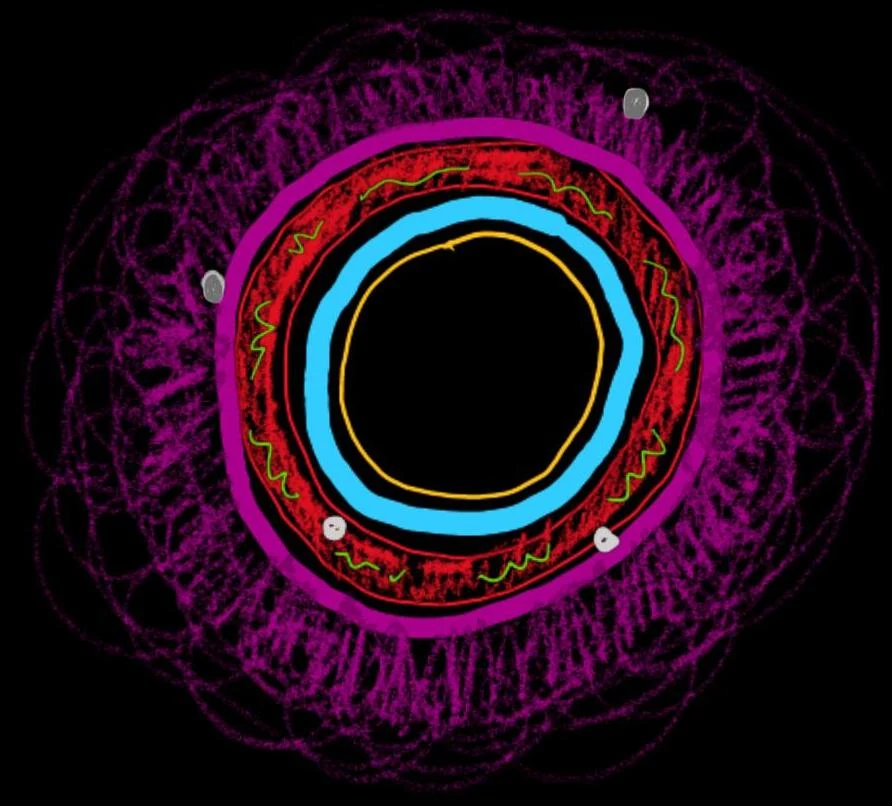
The Vascular Hierarchy: From Arteries to Capillaries
The cardiovascular system is organized into a hierarchy of vessels that manage blood pressure and facilitate material exchange.
Arteries: The Pressure Reservoirs
Arteries carry blood away from the heart. They are categorized based on their size and composition:
- Elastic Arteries: These are the largest arteries, located closest to the heart (e.g., the aorta and pulmonary trunk). They contain high amounts of elastic fibers in the tunica media to handle the high-pressure surges from ventricular contraction.
- Muscular Arteries: Also known as distributing arteries, these are smaller than elastic arteries and deliver blood to specific organs (e.g., brachial or femoral arteries). They have a more prominent tunica media of smooth muscle.
- Arterioles: The smallest arterial vessels, which connect arteries to capillaries. They play a massive role in regulating blood flow into capillary beds.
Capillaries: The Site of Exchange
Capillaries are the tiniest blood vessels and represent the functional unit of the circulatory system. They have the lowest blood flow velocity and the greatest surface area, which is optimized for the exchange of gases, nutrients, and wastes between blood and interstitial fluid. There are three main types:
- Continuous Capillaries: The most common type, featuring a continuous endothelial lining.
- Fenestrated Capillaries: Contain small pores (fenestrations) that allow for faster exchange of small molecules.
- Discontinuous Capillaries (Sinusoids): Have large gaps in the endothelial lining and basement membrane, allowing even large proteins or cells to pass through.
Exam Tip: Microcirculation Control
In a capillary bed, blood flow is regulated by precapillary sphincters—rings of smooth muscle that can contract to divert blood away from certain areas of the tissue.
Veins: The Volume Reservoirs
Veins return blood to the heart. Unlike arteries, veins operate under low pressure and hold the majority of the body's blood volume. To combat gravity and low pressure, veins utilize venous valves to prevent the backflow of blood. Additionally, the skeletal muscle pump—the contraction of surrounding muscles—helps squeeze blood upward through the veins.
Clinical Correlations: Vascular Disorders
Understanding healthy anatomy is vital for recognizing pathology:
- Arteriosclerosis: The hardening and thickening of arterial walls.
- Aneurysm: A dangerous, abnormal bulge or "ballooning" in an artery wall that can rupture.
- Arteriovenous Malformation: An abnormal direct connection between arteries and veins that lacks a capillary bed.
- Diabetic Angiopathy: Damage to blood vessels (often narrowing the lumen) caused by diabetes, which can lead to severe complications like tissue necrosis.
- Deep Vein Thrombosis (DVT): The formation of a blood clot in a deep vein. If this clot breaks loose and travels through the bloodstream, it is called an embolus, which can cause a life-threatening pulmonary embolism.
- Varicose Veins: Occur when venous valves fail, causing blood to pool and veins to become enlarged and twisted.
Hemostasis: The Process of Blood Clotting
When a vessel is injured, the body must prevent excessive blood loss through hemostasis. This involves:
- Platelet Plug Formation: Platelets (small fragments of megakaryocytes) adhere to the site of injury.
- Coagulation: Clotting factors in the plasma trigger a cascade that converts the inactive protein fibrinogen into the active, thread-like protein fibrin.
- Clot Formation: The fibrin strands create a mesh that traps red blood cells and platelets, forming a stable clot.
The Lymphatic System: Fluid Balance and Immunity
The lymphatic system is a one-way system that collects excess interstitial fluid (the fluid surrounding cells) and returns it to the venous circulation.
Lymphatic Structure
- Lymphatic Capillaries: These are closed-ended vessels that collect fluid from the tissues.
- Lymph Nodes: Small, bean-shaped organs that act as filters. They contain lymphocytes (part of the adaptive immune system) that monitor the lymph for pathogens.
- Lymphatic Organs: This includes the thymus (where T-cells mature), the spleen (which filters blood and contains red and white pulp), and the tonsils.
Lymphatic Drainage Pathways
Lymph travels from capillaries through collecting vessels and lymphatic trunks until it reaches one of two main ducts:
- Right Lymphatic Duct: Drains lymph from approximately 1/4 of the body (the right upper quadrant) into the right subclavian vein.
- Thoracic Duct: Drains the remaining 3/4 of the body into the left subclavian vein.
Clinical Note: Lymphedema
When lymphatic vessels are blocked—for example, by a parasitic worm (causing elephantiasis) or surgical removal of nodes—fluid accumulates in the tissues, leading to severe swelling known as lymphedema.