


The knee is a complex hinge joint that relies heavily on the muscular structures of the thigh to maintain stability and facilitate movement. While ligaments like the ACL and MCL provide passive stability, the muscles located on the anterior (front) aspect of the thigh are the primary drivers of knee extension. Understanding these muscles is essential for grasping how the human body manages locomotion, stabilizes the patella (kneecap), and distributes force during weight-bearing activities.
The anterior compartment is dominated by the quadriceps femoris group, a powerful four-headed muscle complex, supplemented by the long, slender sartorius muscle.
The Quadriceps Femoris Group
The quadriceps femoris is the primary extensor of the knee joint. These four muscles converge into a single tendon that encompasses the patella and inserts onto the tibial tuberosity.
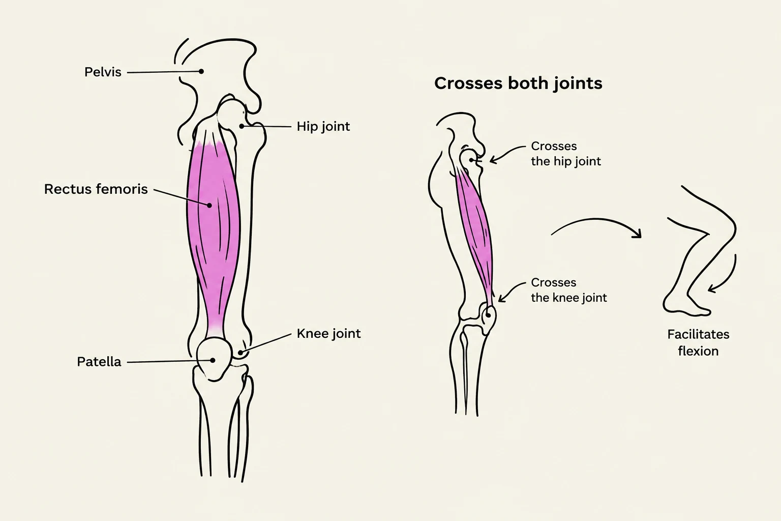
Rectus Femoris
The rectus femoris is the most superficial of the four quadriceps muscles. What distinguishes it from its counterparts is its "biarticular" nature. While the other three heads of the quadriceps only cross the knee joint, the rectus femoris crosses both the hip and the knee joints.
Because it originates on the pelvis (the anterior inferior iliac spine), it can assist in both hip flexion and knee extension. This dual role makes it a critical muscle for complex movements like running or climbing stairs, where both hip and knee actions must be coordinated.
Vastus Lateralis
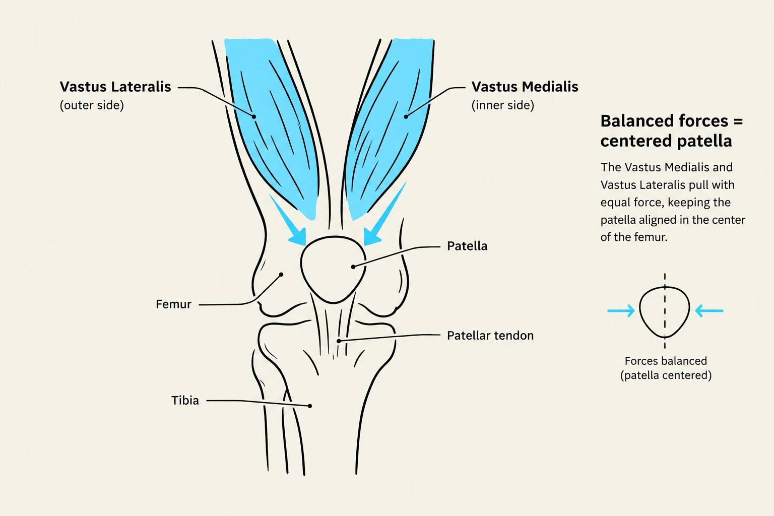
On the outer (lateral) side of the thigh lies the vastus lateralis. This is the largest component of the quadriceps group. It provides significant power for knee extension and contributes to the lateral tension of the patella. In clinical settings, practitioners often look at the Vastus Lateralis Obliquus (VLO), which refers to the specific fiber orientation that pulls the patella laterally.
Keep reading for free.
Create an account to finish this reading and make it part of your study routine.
Continue reading freeAlready have an account? Log in
Updated