An in-depth exploration of the muscles responsible for facial expressions, detailing their anatomy, innervation by the facial nerve, and specific functions in creating various expressions. Covers muscles of the scalp, ears, eyes, nose, mouth, and neck.
The human face is one of our most expressive features, capable of communicating a vast spectrum of emotions ranging from joy and surprise to anger and sorrow. This expressive capability is made possible by a specialized group of muscles located in the scalp, ears, eyes, nose, mouth, and neck. Unlike many other skeletal muscles that attach bone to bone, many muscles of facial expression attach to the skin, allowing them to pull the integument into various shapes and patterns.
Innervation and Control
A critical concept for understanding facial movement is the nerve supply. Most muscles involved in facial expression—including those of the ears, scalp, neck, eyes, nose, and mouth—are innervated by the seventh cranial nerve (CN VII), also known as the facial nerve.
Exam Tip: If a clinical scenario describes a patient unable to smile or close their eyes due to facial paralysis, look for damage to the seventh cranial nerve (Facial Nerve). However, be careful: the muscle that raises the upper eyelid (Levator palpebrae superioris) is actually supplied by the third cranial nerve (Oculomotor nerve).
Muscles of the Ears and Scalp
The muscles of the ear and scalp contribute to subtle movements of the head and ears, often associated with attentiveness or emotional shifts.
The Auricular Muscles
The muscles of the ear are categorized by their position relative to the ear:
Anterior auricular muscles: Arise from the connective tissue of the scalp and run posteriorly to the anterior part of the ear, pulling the ears slightly forward.
Superior auricular muscles: Arise from the connective tissue of the scalp above the ear and run down to insert into the upper part of the ear, serving to raise the ears.
Posterior auricular muscles: Arise from the superior nuchal line and run forward to insert into the posterior part of the ear, pulling the ears backward.
Diagram illustrating the three groups of auricular muscles: anterior, superior, and posterior, and their respective functions in moving the ear.
The Occipitofrontalis (Epicranius)
The occipitofrontalis is a paired muscle consisting of groups of fibers in the front and back, connected by a broad, flat band of fascia. These fibers originate from the connective tissue of the scalp. This muscle is responsible for the movement of the scalp: forward movement can result in a frown or squint, while backward movement raises the skin of the forehead, as seen in expressions of surprise.
Muscles of the Eye and Nose
The eyes and nose are central to facial communication, particularly regarding focus and reaction.
Eye Muscles
The muscles surrounding the eyes control eyelid movement and eyebrow positioning:
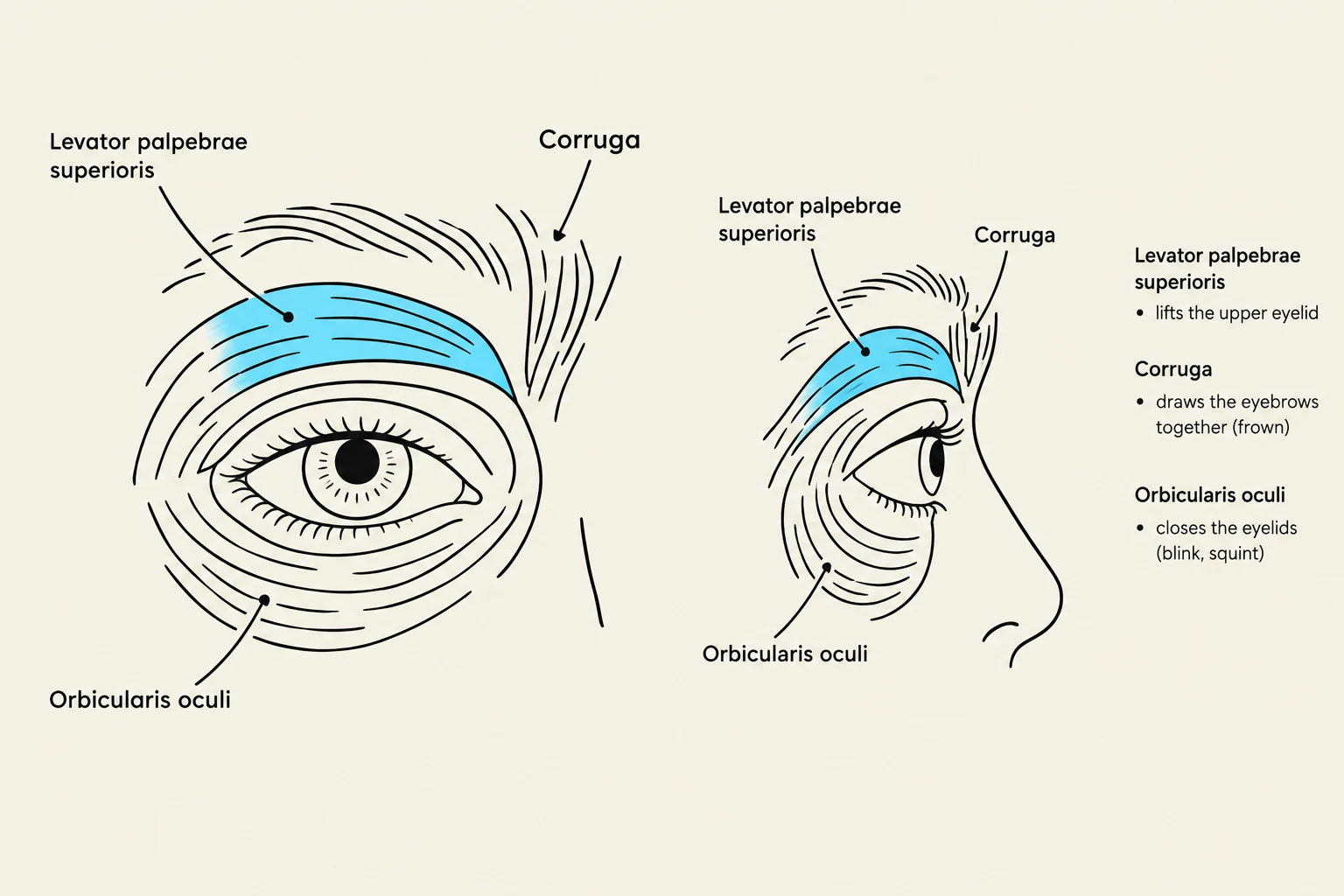
Orbicularis oculi: This muscle encircles the eye. It has two parts: the orbital part (encircling the eye) and the palpebral part (located in the eyelid). It attaches to the skull at the medial and lateral edges of the orbit.
Levator palpebrae superioris: This muscle is responsible for raising the upper eyelid. Note that it is supplied by the oculomotor nerve (CN III), not the facial nerve.
Corrugator: This muscle runs from the bridge of the nose up and lateral to the eyebrow, pulling the eyebrow medially and downward to create a frown.
Procerus: Running from the bridge of the nose up into the medial part of the eyebrow, this muscle pulls the medial end of the eyebrow down, contributing to a squint or frown.
Diagrams illustrating the muscles of the eye, including the levator palpebrae superioris, corruga, and orbicularis oculi, with descriptions of their functions.
Nose Muscles
The muscles of the nose primarily encircle the nostrils. The nasalis muscle is the primary actor here, consisting of two parts: the dilator naris, which flares or dilates the nostrils, and the compressor naris, which causes the nostrils to close or compress.
Muscles of the Mouth and Neck
The muscles of the mouth are perhaps the most complex, as they influence not only expression and speech but also assist in the mechanical process of chewing (mastication).
The Orbicularis Oris and Surrounding Muscles
The orbicularis oris circles the oral cavity within the lip tissue, acting like a "purse string" to close and compress the lips. It has bony attachments at the anterior nasal spine and the midline above the chin.
Several muscles work to move the lips up or down:
Levator labii superioris: Elevates the upper lip.
Zygomaticus minor: A small muscle from the zygomatic bone that also raises the upper lip, though it is poorly developed and exerts little influence.
Depressor labii inferioris: Pulls the lower lip down, often seen in a pout.
Mentalis: Located on the anterior surface of the mandible, it pulls the skin of the chin upward.
Smiling and Frowning
Expressions like smiling or pulling the corners of the mouth are controlled by specific muscles:
Zygomaticus major: Originates from the zygomatic bone and inserts into the orbicularis oris at the angle of the mouth, elevating the corners of the mouth in a smile.
Levator anguli oris: Lies deep to other zygomatic muscles and pulls the angles of the mouth up and toward the midline.
Depressor anguli oris: Pulls the corners of the mouth down.
Risorius: A small muscle that aids in smiling but is usually poorly developed.
The Buccinator and Platysma
The buccinator is considered one of the most important muscles of the mouth. It forms the musculature of the cheek and inserts into the orbicularis oris. It serves as an accessory muscle of mastication by compressing the cheek and pushing food back onto the occlusal surfaces of the teeth during chewing.
Diagram illustrating the Zygomaticus major, Orbicularis oris, and Buccinator muscles of the face and their functions.
Finally, the platysma is a broad, flat muscle of the neck. Its upper fibers attach to the inferior border of the mandible, and it extends down to the skin of the chest below the clavicle. It functions to move the skin of the neck and can pull the corners of the mouth down.
Clinical Note: The normal pressure exerted by mouth muscles helps keep teeth in alignment. However, abnormal pressures—such as those caused by chronic cheek biting or lip compression—can actually cause teeth to move out of their proper alignment.
Keep reading for free.
Create an account to finish this reading and make it part of your study routine.